6CFI

Crystal structure of Rad4-Rad23 bound to a 6-4 photoproduct UV lesion


Experimental Data Snapshot

  • Method: X-RAY DIFFRACTION
  • Resolution: 3.36 Å
  • R-Value Free: 0.268 
  • R-Value Work: 0.219 
  • R-Value Observed: 0.221 

wwPDB Validation   3D Report Full Report


This is version 1.3 of the entry. See complete history


Literature

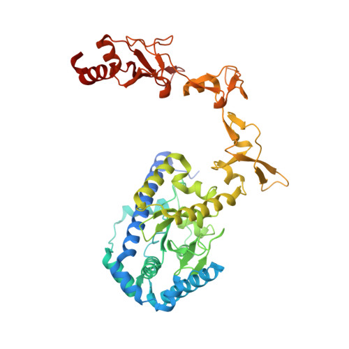
Structure and mechanism of pyrimidine-pyrimidone (6-4) photoproduct recognition by the Rad4/XPC nucleotide excision repair complex.

Paul, D.Mu, H.Zhao, H.Ouerfelli, O.Jeffrey, P.D.Broyde, S.Min, J.H.

(2019) Nucleic Acids Res 47: 6015-6028

  • DOI: https://doi.org/10.1093/nar/gkz359
  • Primary Citation of Related Structures:  
    6CFI

  • PubMed Abstract: 

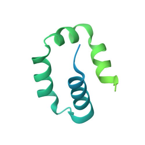
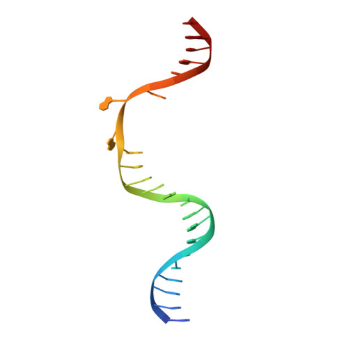
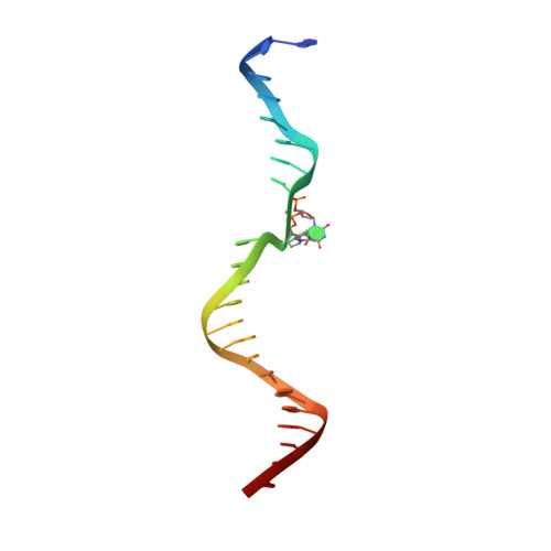
    Failure in repairing ultraviolet radiation-induced DNA damage can lead to mutations and cancer. Among UV-lesions, the pyrimidine-pyrimidone (6-4) photoproduct (6-4PP) is removed from the genome much faster than the cyclobutane pyrimidine dimer (CPD), owing to the more efficient recognition of 6-4PP by XPC-RAD23B, a key initiator of global-genome nucleotide excision repair (NER). Here, we report a crystal structure of a Rad4-Rad23 (yeast XPC-Rad23B ortholog) bound to 6-4PP-containing DNA and 4-μs molecular dynamics (MD) simulations examining the initial binding of Rad4 to 6-4PP or CPD. This first structure of Rad4/XPC bound to a physiological substrate with matched DNA sequence shows that Rad4 flips out both 6-4PP-containing nucleotide pairs, forming an 'open' conformation. The MD trajectories detail how Rad4/XPC initiates 'opening' 6-4PP: Rad4 initially engages BHD2 to bend/untwist DNA from the minor groove, leading to unstacking and extrusion of the 6-4PP:AA nucleotide pairs towards the major groove. The 5' partner adenine first flips out and is captured by a BHD2/3 groove, while the 3' adenine extrudes episodically, facilitating ensuing insertion of the BHD3 β-hairpin to open DNA as in the crystal structure. However, CPD resists such Rad4-induced structural distortions. Untwisting/bending from the minor groove may be a common way to interrogate DNA in NER.


  • Organizational Affiliation

    Department of Chemistry & Biochemistry, Baylor University, Waco, TX 76798, USA.


Macromolecules

Find similar proteins by:  (by identity cutoff)  |  3D Structure
Entity ID: 1
MoleculeChains Sequence LengthOrganismDetailsImage
DNA repair protein RAD4538Saccharomyces cerevisiae S288CMutation(s): 0 
Gene Names: RAD4YER162C
UniProt
Find proteins for P14736 (Saccharomyces cerevisiae (strain ATCC 204508 / S288c))
Explore P14736 
Go to UniProtKB:  P14736
Entity Groups  
Sequence Clusters30% Identity50% Identity70% Identity90% Identity95% Identity100% Identity
UniProt GroupP14736
Sequence Annotations
Expand
  • Reference Sequence
Find similar proteins by:  (by identity cutoff)  |  3D Structure
Entity ID: 2
MoleculeChains Sequence LengthOrganismDetailsImage
UV excision repair protein RAD23B [auth X]171Saccharomyces cerevisiae S288CMutation(s): 0 
Gene Names: RAD23YEL037CSYGP-ORF29
UniProt
Find proteins for P32628 (Saccharomyces cerevisiae (strain ATCC 204508 / S288c))
Explore P32628 
Go to UniProtKB:  P32628
Entity Groups  
Sequence Clusters30% Identity50% Identity70% Identity90% Identity95% Identity100% Identity
UniProt GroupP32628
Sequence Annotations
Expand
  • Reference Sequence

Find similar nucleic acids by:  Sequence   |   3D Structure  

Entity ID: 3
MoleculeChains LengthOrganismImage
DNA('-D(*TP*TP*GP*AP*CP*TP*CP*AP*AP*CP*AP*TP*CP*CP*AP*AP*AP*GP*CP*TP*AP*CP*AP*A)-')C [auth W]24synthetic construct
Sequence Annotations
Expand
  • Reference Sequence

Find similar nucleic acids by:  Sequence   |   3D Structure  

Entity ID: 4
MoleculeChains LengthOrganismImage
DNA (5'-D(*AP*TP*TP*GP*TP*AP*GP*CP*(T64)P*TP*GP*GP*AP*TP*GP*TP*TP*GP*AP*GP*TP*CP*A)-3')D [auth Y]23synthetic construct
Sequence Annotations
Expand
  • Reference Sequence
Experimental Data & Validation

Experimental Data

  • Method: X-RAY DIFFRACTION
  • Resolution: 3.36 Å
  • R-Value Free: 0.268 
  • R-Value Work: 0.219 
  • R-Value Observed: 0.221 
  • Space Group: P 41 21 2
Unit Cell:
Length ( Å )Angle ( ˚ )
a = 80.201α = 90
b = 80.201β = 90
c = 405.025γ = 90
Software Package:
Software NamePurpose
PHENIXrefinement
XDSdata reduction
XDSdata scaling
PHENIXphasing

Structure Validation

View Full Validation Report


Entry History & Funding Information

Deposition Data


Funding OrganizationLocationGrant Number
National Science Foundation (NSF, United States)United StatesMCB-1412692

Revision History  (Full details and data files)

  • Version 1.0: 2019-02-27
    Type: Initial release
  • Version 1.1: 2019-11-27
    Changes: Author supporting evidence
  • Version 1.2: 2021-05-26
    Changes: Database references, Structure summary
  • Version 1.3: 2024-03-13
    Changes: Data collection, Database references