6CCB

Crystal structure of 253-11 SOSIP trimer in complex with 10-1074 Fab


Experimental Data Snapshot

  • Method: X-RAY DIFFRACTION
  • Resolution: 6.50 Å
  • R-Value Free: 0.366 
  • R-Value Work: 0.298 
  • R-Value Observed: 0.305 

wwPDB Validation   3D Report Full Report


Ligand Structure Quality Assessment 


This is version 2.1 of the entry. See complete history


Literature

Molecular Basis of Unusually High Neutralization Resistance in Tier 3 HIV-1 Strain 253-11.

Moyo, T.Ereno-Orbea, J.Jacob, R.A.Pavillet, C.E.Kariuki, S.M.Tangie, E.N.Julien, J.P.Dorfman, J.R.

(2018) J Virol 92

  • DOI: https://doi.org/10.1128/JVI.02261-17
  • Primary Citation of Related Structures:  
    6CCB

  • PubMed Abstract: 

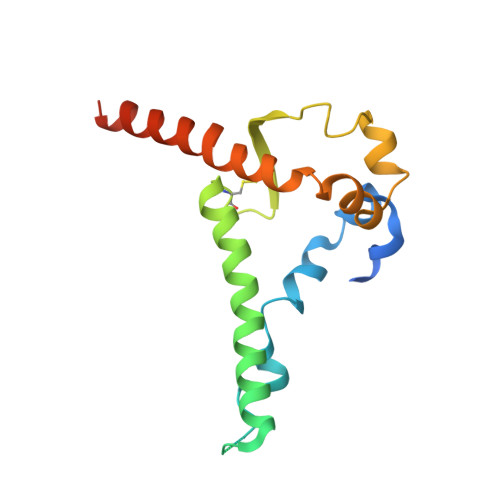
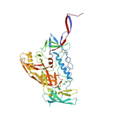
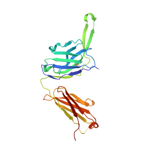
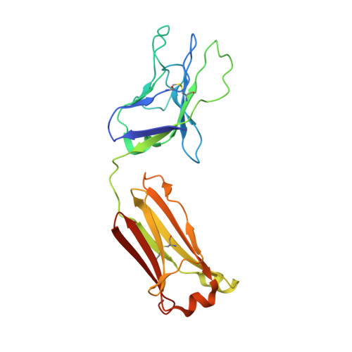
    Understanding the mechanisms used by HIV-1 to evade antibody neutralization may contribute to the design of a high-coverage vaccine. The tier 3 virus 253-11 is poorly neutralized by subtype-matched and subtype C sera, even compared to other tier 3 viruses, and is also recognized poorly by V3/glycan-targeting monoclonal antibodies (MAbs). We found that sequence polymorphisms in the V3 loop and N-linked glycosylation sites contribute only minimally to the high neutralization resistance of 253-11. Interestingly, the 253-11 membrane-proximal external region (MPER) is rarely recognized by sera in the context of the wild-type virus but is commonly recognized in the context of an HIV-2 chimera, suggesting steric or kinetic hindrance of binding to MPER in the native envelope (Env). Mutations in the 253-11 MPER, which were previously reported to increase the lifetime of the prefusion Env conformation, affected the resistance of 253-11 to antibodies targeting various epitopes on HIV-1 Env, presumably destabilizing its otherwise stable, closed trimer structure. To gain insight into the structure of 253-11, we constructed and crystallized a recombinant 253-11 SOSIP trimer. The resulting structure revealed that the heptad repeat helices in gp41 are drawn in close proximity to the trimer axis and that gp120 protomers also showed a relatively compact disposition around the trimer axis. These observations give substantial insight into the molecular features of an envelope spike from a tier 3 virus and into possible mechanisms that may contribute to its unusually high neutralization resistance. IMPORTANCE HIV-1 isolates that are highly resistant to broadly neutralizing antibodies could limit the efficacy of an antibody-based vaccine. We studied 253-11, which is highly resistant to commonly elicited neutralizing antibodies. To further understand its resistance, we made mutations that are known to delay fusion and thus increase the time that the virus spends in the open conformation following CD4 binding. Interestingly, we found that these mutations affect the 253-11 envelope (Env) spike before CD4 binding, presumably by destabilizing the trimer structure. To gain further information about the structure of the 253-11 Env trimer, we generated a recombinant 253-11 SOSIP trimer. The crystal structure of the SOSIP trimer revealed that the gp41 helices and the gp120 protomers were drawn in toward the center of the molecule compared to most solved HIV-1 Env structures. These observations provide insight into the distinct molecular features of a tier 3 envelope spike.


  • Organizational Affiliation

    Division of Immunology, Department of Pathology, University of Cape Town, Cape Town, South Africa.


Macromolecules
Find similar proteins by:  (by identity cutoff)  |  3D Structure
Entity ID: 1
MoleculeChains Sequence LengthOrganismDetailsImage
Glycoprotein 41
A, B
162Human immunodeficiency virus 1Mutation(s): 1 
Gene Names: env
UniProt
Find proteins for B2YFS0 (Human immunodeficiency virus 1)
Explore B2YFS0 
Go to UniProtKB:  B2YFS0
Entity Groups  
Sequence Clusters30% Identity50% Identity70% Identity90% Identity95% Identity100% Identity
UniProt GroupB2YFS0
Sequence Annotations
Expand
  • Reference Sequence
Find similar proteins by:  (by identity cutoff)  |  3D Structure
Entity ID: 2
MoleculeChains Sequence LengthOrganismDetailsImage
Glycoprotein 120C,
F [auth G]
486Human immunodeficiency virus 1Mutation(s): 3 
Gene Names: env
UniProt
Find proteins for B2YFS0 (Human immunodeficiency virus 1)
Explore B2YFS0 
Go to UniProtKB:  B2YFS0
Entity Groups  
Sequence Clusters30% Identity50% Identity70% Identity90% Identity95% Identity100% Identity
UniProt GroupB2YFS0
Sequence Annotations
Expand
  • Reference Sequence
Find similar proteins by:  (by identity cutoff)  |  3D Structure
Entity ID: 3
MoleculeChains Sequence LengthOrganismDetailsImage
10-1074 FAB heavy chainD,
G [auth H]
237Homo sapiensMutation(s): 0 
Entity Groups  
Sequence Clusters30% Identity50% Identity70% Identity90% Identity95% Identity100% Identity
Sequence Annotations
Expand
  • Reference Sequence
Find similar proteins by:  (by identity cutoff)  |  3D Structure
Entity ID: 4
MoleculeChains Sequence LengthOrganismDetailsImage
10-1074 Fab light chainE,
H [auth L]
215Homo sapiensMutation(s): 0 
Entity Groups  
Sequence Clusters30% Identity50% Identity70% Identity90% Identity95% Identity100% Identity
Sequence Annotations
Expand
  • Reference Sequence
Oligosaccharides

Help

Entity ID: 5
MoleculeChains Length2D Diagram Glycosylation3D Interactions
alpha-D-mannopyranose-(1-6)-alpha-D-mannopyranose-(1-6)-[alpha-D-mannopyranose-(1-3)]beta-D-mannopyranose-(1-4)-2-acetamido-2-deoxy-beta-D-glucopyranose-(1-4)-2-acetamido-2-deoxy-beta-D-glucopyranoseI [auth F]6N-Glycosylation
Glycosylation Resources
GlyTouCan:  G34442SS
GlyCosmos:  G34442SS
GlyGen:  G34442SS
Entity ID: 6
MoleculeChains Length2D Diagram Glycosylation3D Interactions
alpha-D-mannopyranose-(1-2)-alpha-D-mannopyranose-(1-2)-alpha-D-mannopyranose-(1-3)-[alpha-D-mannopyranose-(1-2)-alpha-D-mannopyranose-(1-6)-[alpha-D-mannopyranose-(1-3)]alpha-D-mannopyranose-(1-6)]beta-D-mannopyranose-(1-4)-2-acetamido-2-deoxy-beta-D-glucopyranose-(1-4)-2-acetamido-2-deoxy-beta-D-glucopyranoseJ [auth I]10N-Glycosylation
Glycosylation Resources
GlyTouCan:  G40702WU
GlyCosmos:  G40702WU
GlyGen:  G40702WU
Entity ID: 7
MoleculeChains Length2D Diagram Glycosylation3D Interactions
alpha-D-mannopyranose-(1-6)-beta-D-mannopyranose-(1-4)-2-acetamido-2-deoxy-beta-D-glucopyranose-(1-4)-2-acetamido-2-deoxy-beta-D-glucopyranoseK [auth J]4N-Glycosylation
Glycosylation Resources
GlyTouCan:  G22573RC
GlyCosmos:  G22573RC
GlyGen:  G22573RC
Entity ID: 8
MoleculeChains Length2D Diagram Glycosylation3D Interactions
alpha-D-mannopyranose-(1-2)-alpha-D-mannopyranose-(1-3)-[alpha-D-mannopyranose-(1-3)-[alpha-D-mannopyranose-(1-6)]alpha-D-mannopyranose-(1-6)]beta-D-mannopyranose-(1-4)-2-acetamido-2-deoxy-beta-D-glucopyranose-(1-4)-2-acetamido-2-deoxy-beta-D-glucopyranoseL [auth K]8N-Glycosylation
Glycosylation Resources
GlyTouCan:  G80966KZ
GlyCosmos:  G80966KZ
GlyGen:  G80966KZ
Small Molecules
Ligands 1 Unique
IDChains Name / Formula / InChI Key2D Diagram3D Interactions
NAG
Query on NAG

Download Ideal Coordinates CCD File 
AA [auth C]
BA [auth C]
CA [auth C]
DA [auth C]
EA [auth G]
AA [auth C],
BA [auth C],
CA [auth C],
DA [auth C],
EA [auth G],
FA [auth G],
GA [auth G],
HA [auth G],
IA [auth G],
JA [auth G],
KA [auth G],
LA [auth G],
M [auth A],
MA [auth G],
N [auth A],
NA [auth G],
O [auth B],
OA [auth G],
P [auth B],
PA [auth G],
Q [auth C],
QA [auth G],
R [auth C],
RA [auth G],
S [auth C],
T [auth C],
U [auth C],
V [auth C],
W [auth C],
X [auth C],
Y [auth C],
Z [auth C]
2-acetamido-2-deoxy-beta-D-glucopyranose
C8 H15 N O6
OVRNDRQMDRJTHS-FMDGEEDCSA-N
Experimental Data & Validation

Experimental Data

  • Method: X-RAY DIFFRACTION
  • Resolution: 6.50 Å
  • R-Value Free: 0.366 
  • R-Value Work: 0.298 
  • R-Value Observed: 0.305 
  • Space Group: H 3
Unit Cell:
Length ( Å )Angle ( ˚ )
a = 237.284α = 90
b = 237.284β = 90
c = 282.044γ = 120
Software Package:
Software NamePurpose
PHENIXrefinement
XDSdata reduction
XSCALEdata scaling
PHASERphasing

Structure Validation

View Full Validation Report


Ligand Structure Quality Assessment 


Entry History & Funding Information

Deposition Data


Funding OrganizationLocationGrant Number
Canadian Institutes of Health Research (CIHR)CanadaNIH-150414

Revision History  (Full details and data files)

  • Version 1.0: 2018-04-04
    Type: Initial release
  • Version 1.1: 2018-04-18
    Changes: Data collection, Database references
  • Version 1.2: 2018-07-11
    Changes: Data collection, Database references
  • Version 1.3: 2020-01-08
    Changes: Author supporting evidence
  • Version 2.0: 2020-07-29
    Type: Remediation
    Reason: Carbohydrate remediation
    Changes: Atomic model, Data collection, Derived calculations, Structure summary
  • Version 2.1: 2023-10-04
    Changes: Data collection, Database references, Derived calculations, Refinement description, Structure summary