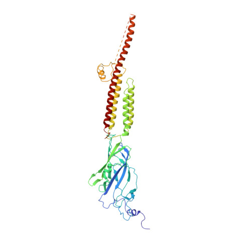
6BE1

Cryo-EM structure of serotonin receptor


Experimental Data Snapshot

  • Method: ELECTRON MICROSCOPY
  • Resolution: 4.31 Å
  • Aggregation State: PARTICLE 
  • Reconstruction Method: SINGLE PARTICLE 

wwPDB Validation   3D Report Full Report


This is version 2.0 of the entry. See complete history


Literature

Cryo-EM structure of 5-HT

Basak, S.Gicheru, Y.Samanta, A.Molugu, S.K.Huang, W.Fuente, M.Hughes, T.Taylor, D.J.Nieman, M.T.Moiseenkova-Bell, V.Chakrapani, S.

(2018) Nat Commun 9: 514-514

  • DOI: https://doi.org/10.1038/s41467-018-02997-4
  • Primary Citation of Related Structures:  
    6BE1

  • PubMed Abstract: 

    Serotonin receptors (5-HT 3A R) directly regulate gut movement, and drugs that inhibit 5-HT 3A R function are used to control emetic reflexes associated with gastrointestinal pathologies and cancer therapies. The 5-HT 3A R function involves a finely tuned orchestration of three domain movements that include the ligand-binding domain, the pore domain, and the intracellular domain. Here, we present the structure from the full-length 5-HT 3A R channel in the apo-state determined by single-particle cryo-electron microscopy at a nominal resolution of 4.3 Å. In this conformation, the ligand-binding domain adopts a conformation reminiscent of the unliganded state with the pore domain captured in a closed conformation. In comparison to the 5-HT 3A R crystal structure, the full-length channel in the apo-conformation adopts a more expanded conformation of all the three domains with a characteristic twist that is implicated in gating.


  • Organizational Affiliation

    Department of Physiology and Biophysics, Case Western Reserve University, Cleveland, OH, 44106-4970, USA.


Macromolecules
Find similar proteins by:  (by identity cutoff)  |  3D Structure
Entity ID: 1
MoleculeChains Sequence LengthOrganismDetailsImage
5-hydroxytryptamine receptor 3A
A, B, C, D, E
457Mus musculusMutation(s): 0 
Gene Names: Htr3a
Membrane Entity: Yes 
UniProt & NIH Common Fund Data Resources
Find proteins for P23979 (Mus musculus)
Explore P23979 
Go to UniProtKB:  P23979
IMPC:  MGI:96282
Entity Groups  
Sequence Clusters30% Identity50% Identity70% Identity90% Identity95% Identity100% Identity
UniProt GroupP23979
Sequence Annotations
Expand
  • Reference Sequence
Oligosaccharides

Help

Entity ID: 2
MoleculeChains Length2D Diagram Glycosylation3D Interactions
beta-D-mannopyranose-(1-4)-2-acetamido-2-deoxy-beta-D-glucopyranose-(1-4)-2-acetamido-2-deoxy-beta-D-glucopyranose
F, K
3N-Glycosylation
Glycosylation Resources
GlyTouCan:  G15407YE
GlyCosmos:  G15407YE
GlyGen:  G15407YE
Entity ID: 3
MoleculeChains Length2D Diagram Glycosylation3D Interactions
2-acetamido-2-deoxy-beta-D-glucopyranose-(1-4)-2-acetamido-2-deoxy-beta-D-glucopyranose
G, I, J, L, N
G, I, J, L, N, O, P, Q
2N-Glycosylation
Glycosylation Resources
GlyTouCan:  G42666HT
GlyCosmos:  G42666HT
GlyGen:  G42666HT
Entity ID: 4
MoleculeChains Length2D Diagram Glycosylation3D Interactions
beta-D-mannopyranose-(1-4)-2-acetamido-2-deoxy-beta-D-glucopyranose-(1-3)-2-acetamido-2-deoxy-beta-D-glucopyranose
H
3N/A
Glycosylation Resources
GlyTouCan:  G53081KU
GlyCosmos:  G53081KU
GlyGen:  G53081KU
Entity ID: 5
MoleculeChains Length2D Diagram Glycosylation3D Interactions
beta-D-mannopyranose-(1-3)-2-acetamido-2-deoxy-beta-D-glucopyranose-(1-4)-2-acetamido-2-deoxy-beta-D-glucopyranose
M
3N-Glycosylation
Glycosylation Resources
GlyTouCan:  G44266EX
GlyCosmos:  G44266EX
GlyGen:  G44266EX
Small Molecules
Ligands 5 Unique
IDChains Name / Formula / InChI Key2D Diagram3D Interactions
PX4
Query on PX4

Download Ideal Coordinates CCD File 
BA [auth D],
DA [auth E],
U [auth A],
V [auth A],
Z [auth C]
1,2-DIMYRISTOYL-SN-GLYCERO-3-PHOSPHOCHOLINE
C36 H73 N O8 P
CITHEXJVPOWHKC-UUWRZZSWSA-O
NAG
Query on NAG

Download Ideal Coordinates CCD File 
AA [auth D],
R [auth A],
S [auth A],
W [auth C],
X [auth C]
2-acetamido-2-deoxy-beta-D-glucopyranose
C8 H15 N O6
OVRNDRQMDRJTHS-FMDGEEDCSA-N
BMA
Query on BMA

Download Ideal Coordinates CCD File 
CA [auth E]beta-D-mannopyranose
C6 H12 O6
WQZGKKKJIJFFOK-RWOPYEJCSA-N
CL
Query on CL

Download Ideal Coordinates CCD File 
Y [auth C]CHLORIDE ION
Cl
VEXZGXHMUGYJMC-UHFFFAOYSA-M
NA
Query on NA

Download Ideal Coordinates CCD File 
T [auth A]SODIUM ION
Na
FKNQFGJONOIPTF-UHFFFAOYSA-N
Experimental Data & Validation

Experimental Data

  • Method: ELECTRON MICROSCOPY
  • Resolution: 4.31 Å
  • Aggregation State: PARTICLE 
  • Reconstruction Method: SINGLE PARTICLE 
EM Software:
TaskSoftware PackageVersion
MODEL REFINEMENTPHENIX1.12.2829
RECONSTRUCTIONRELION2.03

Structure Validation

View Full Validation Report


Entry History & Funding Information

Deposition Data


Funding OrganizationLocationGrant Number
National Institutes of Health/National Institute of General Medical Sciences (NIH/NIGMS)United States1R01GM108921
National Institutes of Health/National Institute of General Medical Sciences (NIH/NIGMS)United States3R01GM108921-03S1
American Heart AssociationUnited States17POST33671152

Revision History  (Full details and data files)

  • Version 1.0: 2018-02-07
    Type: Initial release
  • Version 1.1: 2018-02-14
    Changes: Author supporting evidence
  • Version 1.2: 2018-02-28
    Changes: Database references
  • Version 1.3: 2019-12-18
    Changes: Advisory, Author supporting evidence, Derived calculations, Other
  • Version 2.0: 2020-07-29
    Type: Remediation
    Reason: Carbohydrate remediation
    Changes: Advisory, Atomic model, Data collection, Derived calculations, Structure summary