6A88

Crystal Structure of T. gondii prolyl tRNA synthetase with Febrifugine and ATP Analog


Experimental Data Snapshot

  • Method: X-RAY DIFFRACTION
  • Resolution: 2.60 Å
  • R-Value Free: 0.220 
  • R-Value Work: 0.172 
  • R-Value Observed: 0.175 

wwPDB Validation   3D Report Full Report


Ligand Structure Quality Assessment 


This is version 1.2 of the entry. See complete history


Literature

Conformational heterogeneity in apo and drug-bound structures of Toxoplasma gondii prolyl-tRNA synthetase.

Mishra, S.Malhotra, N.Kumari, S.Sato, M.Kikuchi, H.Yogavel, M.Sharma, A.

(2019) Acta Crystallogr F Struct Biol Commun 75: 714-724

  • DOI: https://doi.org/10.1107/S2053230X19014808
  • Primary Citation of Related Structures:  
    6A88, 6AA0

  • PubMed Abstract: 

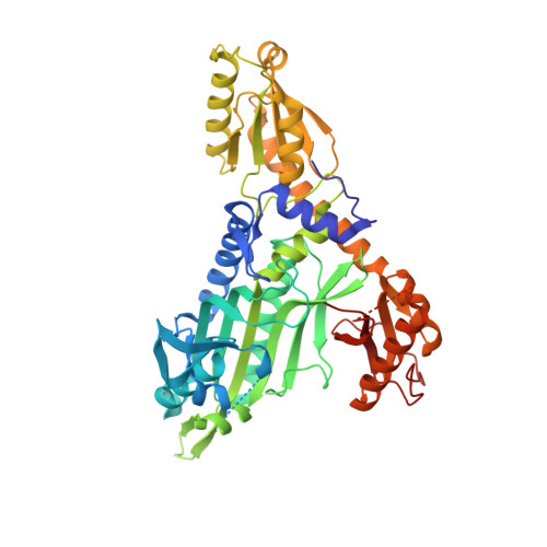
    Prolyl-tRNA synthetase (PRS) is a member of the aminoacyl-tRNA synthetase family that drives protein translation in cells. The apicomplexan PRSs are validated targets of febrifugine (FF) and its halogenated derivative halofuginone (HF). PRSs are of great interest for drug development against Plasmodium falciparum and Toxoplasma gondii. In this study, structures of apo and FF-bound T. gondii (TgPRS) are revealed and the dynamic nature of the conformational changes that occur upon FF binding is unraveled. In addition, this study highlights significant conformational plasticity within two different crystal structures of apo PRSs but not within drug-bound PRSs. The apo PRSs exist in multi-conformational states and manifest pseudo-dimeric structures. In contrast, when FF is bound the PRS dimer adopts a highly symmetrical architecture. It is shown that TgPRS does not display extant fold switching, in contrast to P. falciparum PRS, despite having over 65% sequence identity. Finally, structure-comparison analyses suggest the utility of r.m.s.d. per residue (r.m.s.d. /res ) as a robust tool to detect structural alterations even when the r.m.s.d. is low. Apo TgPRS reveals FF/HF-induced rigidity and this work has implications for drug-design studies that rely on the apo structures of target proteins.


  • Organizational Affiliation

    Structural Parasitology, International Centre for Genetic Engineering and Biotechnology, New Delhi, Aruna Asaf Ali Marg, New Delhi, Delhi 110067, India.


Macromolecules
Find similar proteins by:  (by identity cutoff)  |  3D Structure
Entity ID: 1
MoleculeChains Sequence LengthOrganismDetailsImage
Prolyl-tRNA synthetase (ProRS)
A, B, C, D
500Toxoplasma gondii ME49Mutation(s): 0 
Gene Names: TGME49_219850
EC: 6.1.1.15
UniProt
Find proteins for S8G8I1 (Toxoplasma gondii (strain ATCC 50611 / Me49))
Explore S8G8I1 
Go to UniProtKB:  S8G8I1
Entity Groups  
Sequence Clusters30% Identity50% Identity70% Identity90% Identity95% Identity100% Identity
UniProt GroupS8G8I1
Sequence Annotations
Expand
  • Reference Sequence
Small Molecules
Ligands 3 Unique
IDChains Name / Formula / InChI Key2D Diagram3D Interactions
ANP
Query on ANP

Download Ideal Coordinates CCD File 
E [auth A],
H [auth B],
K [auth C],
N [auth D]
PHOSPHOAMINOPHOSPHONIC ACID-ADENYLATE ESTER
C10 H17 N6 O12 P3
PVKSNHVPLWYQGJ-KQYNXXCUSA-N
9SF (Subject of Investigation/LOI)
Query on 9SF

Download Ideal Coordinates CCD File 
G [auth A],
J [auth B],
M [auth C],
P [auth D]
3-{3-[(2R,3S)-3-hydroxypiperidin-2-yl]-2-oxopropyl}quinazolin-4(3H)-one
C16 H19 N3 O3
FWVHWDSCPKXMDB-CABCVRRESA-N
MG
Query on MG

Download Ideal Coordinates CCD File 
F [auth A],
I [auth B],
L [auth C],
O [auth D]
MAGNESIUM ION
Mg
JLVVSXFLKOJNIY-UHFFFAOYSA-N
Experimental Data & Validation

Experimental Data

  • Method: X-RAY DIFFRACTION
  • Resolution: 2.60 Å
  • R-Value Free: 0.220 
  • R-Value Work: 0.172 
  • R-Value Observed: 0.175 
  • Space Group: P 1
Unit Cell:
Length ( Å )Angle ( ˚ )
a = 76.91α = 89.91
b = 90.882β = 80.3
c = 93.028γ = 75.58
Software Package:
Software NamePurpose
PHENIXrefinement
PHASERphasing
PDB_EXTRACTdata extraction
DIALSdata reduction
DIALSdata scaling

Structure Validation

View Full Validation Report


Ligand Structure Quality Assessment 


Entry History 

Deposition Data

Revision History  (Full details and data files)

  • Version 1.0: 2019-10-23
    Type: Initial release
  • Version 1.1: 2019-11-20
    Changes: Database references
  • Version 1.2: 2023-11-22
    Changes: Data collection, Database references, Derived calculations, Refinement description