6ZWK

Crystal structure of the phosphorylated C-terminal tail of histone H2AX in complex with a specific nanobody (C6 gammaXbody)


Experimental Data Snapshot

  • Method: X-RAY DIFFRACTION
  • Resolution: 1.55 Å
  • R-Value Free: 0.184 
  • R-Value Work: 0.149 
  • R-Value Observed: 0.151 

wwPDB Validation   3D Report Full Report


This is version 1.2 of the entry. See complete history


Literature

A Novel Nanobody Precisely Visualizes Phosphorylated Histone H2AX in Living Cancer Cells under Drug-Induced Replication Stress.

Moeglin, E.Desplancq, D.Stoessel, A.Massute, C.Ranniger, J.McEwen, A.G.Zeder-Lutz, G.Oulad-Abdelghani, M.Chiper, M.Lafaye, P.Di Ventura, B.Didier, P.Poterszman, A.Weiss, E.

(2021) Cancers (Basel) 13

  • DOI: https://doi.org/10.3390/cancers13133317
  • Primary Citation of Related Structures:  
    6ZWK

  • PubMed Abstract: 

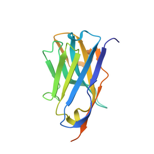
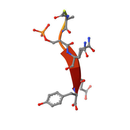
    Histone H2AX phosphorylated at serine 139 (γ-H2AX) is a hallmark of DNA damage, signaling the presence of DNA double-strand breaks and global replication stress in mammalian cells. While γ-H2AX can be visualized with antibodies in fixed cells, its detection in living cells was so far not possible. Here, we used immune libraries and phage display to isolate nanobodies that specifically bind to γ-H2AX. We solved the crystal structure of the most soluble nanobody in complex with the phosphopeptide corresponding to the C-terminus of γ-H2AX and show the atomic constituents behind its specificity. We engineered a bivalent version of this nanobody and show that bivalency is essential to quantitatively visualize γ-H2AX in fixed drug-treated cells. After labelling with a chemical fluorophore, we were able to detect γ-H2AX in a single-step assay with the same sensitivity as with validated antibodies. Moreover, we produced fluorescent nanobody-dTomato fusion proteins and applied a transduction strategy to visualize with precision γ-H2AX foci present in intact living cells following drug treatment. Together, this novel tool allows performing fast screenings of genotoxic drugs and enables to study the dynamics of this particular chromatin modification in individual cancer cells under a variety of conditions.


  • Organizational Affiliation

    Biotechnologie et Signalisation Cellulaire, UMR 7242, CNRS/Université de Strasbourg, Boulevard S. Brant, 67412 Illkirch, France.


Macromolecules
Find similar proteins by:  (by identity cutoff)  |  3D Structure
Entity ID: 1
MoleculeChains Sequence LengthOrganismDetailsImage
gammaXbody
A, B, C, D, E
A, B, C, D, E, F
150Vicugna pacosMutation(s): 0 
Entity Groups  
Sequence Clusters30% Identity50% Identity70% Identity90% Identity95% Identity100% Identity
Sequence Annotations
Expand
  • Reference Sequence

Find similar proteins by:  Sequence   |   3D Structure  

Entity ID: 2
MoleculeChains Sequence LengthOrganismDetailsImage
Histone H2AX
G, H, I, J, K
G, H, I, J, K, L
10Homo sapiensMutation(s): 1 
UniProt & NIH Common Fund Data Resources
Find proteins for P16104 (Homo sapiens)
Explore P16104 
Go to UniProtKB:  P16104
PHAROS:  P16104
GTEx:  ENSG00000188486 
Entity Groups  
Sequence Clusters30% Identity50% Identity70% Identity90% Identity95% Identity100% Identity
UniProt GroupP16104
Sequence Annotations
Expand
  • Reference Sequence
Small Molecules
Modified Residues  1 Unique
IDChains TypeFormula2D DiagramParent
SEP
Query on SEP
G, H, I, J, K
G, H, I, J, K, L
L-PEPTIDE LINKINGC3 H8 N O6 PSER
Experimental Data & Validation

Experimental Data

  • Method: X-RAY DIFFRACTION
  • Resolution: 1.55 Å
  • R-Value Free: 0.184 
  • R-Value Work: 0.149 
  • R-Value Observed: 0.151 
  • Space Group: P 31
Unit Cell:
Length ( Å )Angle ( ˚ )
a = 87.412α = 90
b = 87.412β = 90
c = 105.409γ = 120
Software Package:
Software NamePurpose
XDSdata reduction
Aimlessdata scaling
PHASERphasing
PHENIXrefinement
PDB_EXTRACTdata extraction

Structure Validation

View Full Validation Report


Entry History & Funding Information

Deposition Data


Funding OrganizationLocationGrant Number
French National Research AgencyFranceFRISBI ANR-10-INBS-05

Revision History  (Full details and data files)

  • Version 1.0: 2021-07-21
    Type: Initial release
  • Version 1.1: 2021-07-28
    Changes: Database references
  • Version 1.2: 2024-01-31
    Changes: Data collection, Database references, Refinement description