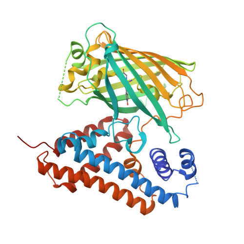
The lack of tools to monitor the dynamics of (pseudo)hypohalous acids in live cells and tissues hinders a better understanding of inflammatory processes. Here we present a fluorescent genetically encoded biosensor, Hypocrates, for the visualization of (pseudo)hypohalous acids and their derivatives. Hypocrates consists of a circularly permuted yellow fluorescent protein integrated into the structure of the transcription repressor NemR from Escherichia coli. We show that Hypocrates is ratiometric, reversible, and responds to its analytes in the 10 6 M -1 s -1 range. Solving the Hypocrates X-ray structure provided insights into its sensing mechanism, allowing determination of the spatial organization in this circularly permuted fluorescent protein-based redox probe. We exemplify its applicability by imaging hypohalous stress in bacteria phagocytosed by primary neutrophils. Finally, we demonstrate that Hypocrates can be utilized in combination with HyPerRed for the simultaneous visualization of (pseudo)hypohalous acids and hydrogen peroxide dynamics in a zebrafish tail fin injury model.
Organizational Affiliation:
Shemyakin-Ovchinnikov Institute of Bioorganic Chemistry, 117997, Moscow, Russia.
Center for Precision Genome Editing and Genetic Technologies for Biomedicine, Pirogov Russian National Research Medical University, 117997, Moscow, Russia.
Laboratory of Experimental Oncology, Pirogov Russian National Research Medical University, 117997, Moscow, Russia.
VIB-VUB Center for Structural Biology, Vlaams Instituut voor Biotechnologie, B-1050, Brussels, Belgium.
Brussels Center for Redox Biology, Vrije Universiteit Brussel, B-1050, Brussels, Belgium.
Shemyakin-Ovchinnikov Institute of Bioorganic Chemistry, 117997, Moscow, Russia. d.s.bilan@gmail.com.
Center for Precision Genome Editing and Genetic Technologies for Biomedicine, Pirogov Russian National Research Medical University, 117997, Moscow, Russia. d.s.bilan@gmail.com.
Laboratory of Experimental Oncology, Pirogov Russian National Research Medical University, 117997, Moscow, Russia. d.s.bilan@gmail.com.
Shemyakin-Ovchinnikov Institute of Bioorganic Chemistry, 117997, Moscow, Russia. belousov@fccps.ru.
Center for Precision Genome Editing and Genetic Technologies for Biomedicine, Pirogov Russian National Research Medical University, 117997, Moscow, Russia. belousov@fccps.ru.
Laboratory of Experimental Oncology, Pirogov Russian National Research Medical University, 117997, Moscow, Russia. belousov@fccps.ru.
Federal Center of Brain Research and Neurotechnologies, Federal Medical Biological Agency, 117997, Moscow, Russia. belousov@fccps.ru.