6ZSM

Crystal structure of rsGCaMP double mutant Ile80His/Val116Ile in the ON state (non-illuminated)


Experimental Data Snapshot

  • Method: X-RAY DIFFRACTION
  • Resolution: 1.95 Å
  • R-Value Free: 0.198 
  • R-Value Work: 0.157 
  • R-Value Observed: 0.159 

wwPDB Validation   3D Report Full Report


This is version 1.3 of the entry. See complete history


Literature

Genetically encoded photo-switchable molecular sensors for optoacoustic and super-resolution imaging.

Mishra, K.Fuenzalida-Werner, J.P.Pennacchietti, F.Janowski, R.Chmyrov, A.Huang, Y.Zakian, C.Klemm, U.Testa, I.Niessing, D.Ntziachristos, V.Stiel, A.C.

(2022) Nat Biotechnol 40: 598-605

  • DOI: https://doi.org/10.1038/s41587-021-01100-5
  • Primary Citation of Related Structures:  
    6TV7, 6YA9, 6ZSM, 6ZSN, 7AUG

  • PubMed Abstract: 

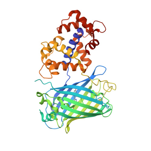
    Reversibly photo-switchable proteins are essential for many super-resolution fluorescence microscopic and optoacoustic imaging methods. However, they have yet to be used as sensors that measure the distribution of specific analytes at the nanoscale or in the tissues of live animals. Here we constructed the prototype of a photo-switchable Ca 2+ sensor based on GCaMP5G that can be switched with 405/488-nm light and describe its molecular mechanisms at the structural level, including the importance of the interaction of the core barrel structure of the fluorescent protein with the Ca 2+ receptor moiety. We demonstrate super-resolution imaging of Ca 2+ concentration in cultured cells and optoacoustic Ca 2+ imaging in implanted tumor cells in mice under controlled Ca 2+ conditions. Finally, we show the generalizability of the concept by constructing examples of photo-switching maltose and dopamine sensors based on periplasmatic binding protein and G-protein-coupled receptor-based sensors.


  • Organizational Affiliation

    Institute of Biological and Medical Imaging, Helmholtz Zentrum München, Neuherberg, Germany.


Macromolecules
Find similar proteins by:  (by identity cutoff)  |  3D Structure
Entity ID: 1
MoleculeChains Sequence LengthOrganismDetailsImage
Green fluorescent protein,Green fluorescent protein,Calmodulin418Aequorea victoriaEntacmaea quadricolorMutation(s): 0 
Gene Names: GFP
UniProt
Find proteins for P42212 (Aequorea victoria)
Explore P42212 
Go to UniProtKB:  P42212
Find proteins for K4DIE3 (Entacmaea quadricolor)
Explore K4DIE3 
Go to UniProtKB:  K4DIE3
Entity Groups  
Sequence Clusters30% Identity50% Identity70% Identity90% Identity95% Identity100% Identity
UniProt GroupsK4DIE3P42212
Sequence Annotations
Expand
  • Reference Sequence
Small Molecules
Ligands 3 Unique
IDChains Name / Formula / InChI Key2D Diagram3D Interactions
EDO
Query on EDO

Download Ideal Coordinates CCD File 
U [auth A],
V [auth A],
W [auth A],
X [auth A],
Y [auth A]
1,2-ETHANEDIOL
C2 H6 O2
LYCAIKOWRPUZTN-UHFFFAOYSA-N
FMT
Query on FMT

Download Ideal Coordinates CCD File 
G [auth A]
H [auth A]
I [auth A]
J [auth A]
K [auth A]
G [auth A],
H [auth A],
I [auth A],
J [auth A],
K [auth A],
L [auth A],
M [auth A],
N [auth A],
O [auth A],
P [auth A],
Q [auth A],
R [auth A],
S [auth A],
T [auth A]
FORMIC ACID
C H2 O2
BDAGIHXWWSANSR-UHFFFAOYSA-N
CA
Query on CA

Download Ideal Coordinates CCD File 
B [auth A],
C [auth A],
D [auth A],
E [auth A],
F [auth A]
CALCIUM ION
Ca
BHPQYMZQTOCNFJ-UHFFFAOYSA-N
Modified Residues  1 Unique
IDChains TypeFormula2D DiagramParent
PIA
Query on PIA
A
L-PEPTIDE LINKINGC14 H15 N3 O4ALA, TYR, GLY
Experimental Data & Validation

Experimental Data

  • Method: X-RAY DIFFRACTION
  • Resolution: 1.95 Å
  • R-Value Free: 0.198 
  • R-Value Work: 0.157 
  • R-Value Observed: 0.159 
  • Space Group: P 41 21 2
Unit Cell:
Length ( Å )Angle ( ˚ )
a = 121.54α = 90
b = 121.54β = 90
c = 96.96γ = 90
Software Package:
Software NamePurpose
REFMACrefinement
PDB_EXTRACTdata extraction
XDSdata reduction
SCALAdata scaling
MOLREPphasing

Structure Validation

View Full Validation Report


Entry History & Funding Information

Deposition Data


Funding OrganizationLocationGrant Number
German Research Foundation (DFG)GermanySTI 656/1-1

Revision History  (Full details and data files)

  • Version 1.0: 2021-10-27
    Type: Initial release
  • Version 1.1: 2021-12-22
    Changes: Database references
  • Version 1.2: 2022-04-27
    Changes: Database references
  • Version 1.3: 2024-01-31
    Changes: Data collection, Refinement description