6ZRK

Crystal structure of H8 haemagglutinin


Experimental Data Snapshot

  • Method: X-RAY DIFFRACTION
  • Resolution: 2.00 Å
  • R-Value Free: 0.257 
  • R-Value Work: 0.224 
  • R-Value Observed: 0.226 

wwPDB Validation   3D Report Full Report


Ligand Structure Quality Assessment 


This is version 1.4 of the entry. See complete history


Literature

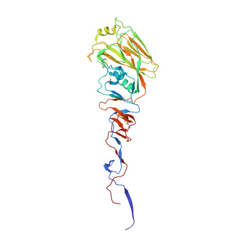
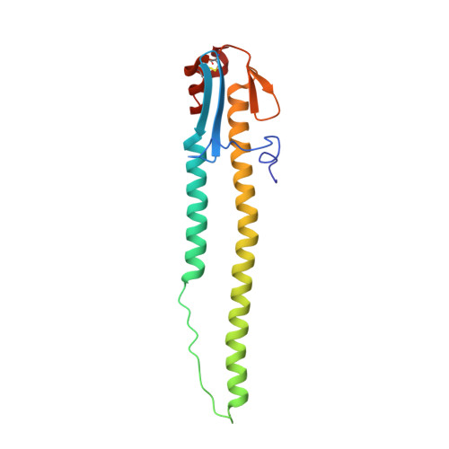
Hemagglutinin Structure and Activities.

Gamblin, S.J.Vachieri, S.G.Xiong, X.Zhang, J.Martin, S.R.Skehel, J.J.

(2021) Cold Spring Harb Perspect Med 11

  • DOI: https://doi.org/10.1101/cshperspect.a038638
  • Primary Citation of Related Structures:  
    6ZRK, 7A9D

  • PubMed Abstract: 

    Hemagglutinins (HAs) are the receptor-binding and membrane fusion glycoproteins of influenza viruses. They recognize sialic acid-containing, cell-surface glycoconjugates as receptors but have limited affinity for them, and, as a consequence, virus attachment to cells requires their interaction with several virus HAs. Receptor-bound virus is transferred into endosomes where membrane fusion by HAs is activated at pH between 5 and 6.5, depending on the strain of virus. Fusion activity requires extensive rearrangements in HA conformation that include extrusion of a buried "fusion peptide" to connect with the endosomal membrane, form a bridge to the virus membrane, and eventually bring both membranes close together. In this review, we give an overview of the structures of the 16 genetically and antigenically distinct subtypes of influenza A HA in relation to these two functions in virus replication and in relation to recognition of HA by antibodies that neutralize infection.


  • Organizational Affiliation

    Structural Biology of Disease Processes Laboratory, The Francis Crick Institute, London NW1 1AT, United Kingdom.


Macromolecules
Find similar proteins by:  (by identity cutoff)  |  3D Structure
Entity ID: 1
MoleculeChains Sequence LengthOrganismDetailsImage
HemagglutininA [auth C]328Influenza A virus (A/blue-winged teal/Guatemala/CIP049-14/2010(H8N4))Mutation(s): 0 
Gene Names: HA
UniProt
Find proteins for G0KTJ4 (Influenza A virus)
Explore G0KTJ4 
Go to UniProtKB:  G0KTJ4
Entity Groups  
Sequence Clusters30% Identity50% Identity70% Identity90% Identity95% Identity100% Identity
UniProt GroupG0KTJ4
Sequence Annotations
Expand
  • Reference Sequence
Find similar proteins by:  (by identity cutoff)  |  3D Structure
Entity ID: 2
MoleculeChains Sequence LengthOrganismDetailsImage
HemagglutininB [auth D]162Influenza A virusMutation(s): 0 
Gene Names: HA
UniProt
Find proteins for G0KTD9 (Influenza A virus)
Explore G0KTD9 
Go to UniProtKB:  G0KTD9
Entity Groups  
Sequence Clusters30% Identity50% Identity70% Identity90% Identity95% Identity100% Identity
UniProt GroupG0KTD9
Sequence Annotations
Expand
  • Reference Sequence
Oligosaccharides

Help

Entity ID: 3
MoleculeChains Length2D Diagram Glycosylation3D Interactions
alpha-D-mannopyranose-(1-3)-[alpha-D-mannopyranose-(1-6)]beta-D-mannopyranose-(1-4)-2-acetamido-2-deoxy-beta-D-glucopyranose-(1-4)-[alpha-L-fucopyranose-(1-6)]2-acetamido-2-deoxy-beta-D-glucopyranoseC [auth A]6N-Glycosylation
Glycosylation Resources
GlyTouCan:  G82348BZ
GlyCosmos:  G82348BZ
GlyGen:  G82348BZ
Entity ID: 4
MoleculeChains Length2D Diagram Glycosylation3D Interactions
2-acetamido-2-deoxy-beta-D-glucopyranose-(1-4)-2-acetamido-2-deoxy-beta-D-glucopyranoseD [auth B],
E,
F
2N-Glycosylation
Glycosylation Resources
GlyTouCan:  G42666HT
GlyCosmos:  G42666HT
GlyGen:  G42666HT
Small Molecules
Ligands 1 Unique
IDChains Name / Formula / InChI Key2D Diagram3D Interactions
NAG
Query on NAG

Download Ideal Coordinates CCD File 
G [auth C]2-acetamido-2-deoxy-beta-D-glucopyranose
C8 H15 N O6
OVRNDRQMDRJTHS-FMDGEEDCSA-N
Experimental Data & Validation

Experimental Data

  • Method: X-RAY DIFFRACTION
  • Resolution: 2.00 Å
  • R-Value Free: 0.257 
  • R-Value Work: 0.224 
  • R-Value Observed: 0.226 
  • Space Group: H 3 2
Unit Cell:
Length ( Å )Angle ( ˚ )
a = 98.739α = 90
b = 98.739β = 90
c = 350.383γ = 120
Software Package:
Software NamePurpose
REFMACrefinement
PDB_EXTRACTdata extraction
SCALAdata scaling
PHASERphasing
xia2data reduction

Structure Validation

View Full Validation Report


Ligand Structure Quality Assessment 


Entry History & Funding Information

Deposition Data


Funding OrganizationLocationGrant Number
The Francis Crick InstituteUnited Kingdom--

Revision History  (Full details and data files)

  • Version 1.0: 2020-12-02
    Type: Initial release
  • Version 1.1: 2020-12-23
    Changes: Derived calculations
  • Version 1.2: 2021-06-16
    Changes: Database references
  • Version 1.3: 2021-10-13
    Changes: Data collection, Database references
  • Version 1.4: 2024-01-31
    Changes: Data collection, Refinement description