6ZPN | pdb_00006zpn

Crystal structure of Chaetomium thermophilum Raptor


Experimental Data Snapshot

  • Method: X-RAY DIFFRACTION
  • Resolution: 3.50 Å
  • R-Value Free: 
    0.245 (Depositor), 0.244 (DCC) 
  • R-Value Work: 
    0.228 (Depositor), 0.228 (DCC) 
  • R-Value Observed: 
    0.229 (Depositor) 

Starting Model: experimental
View more details

wwPDB Validation 3D Report Full Report

Validation slider image for 6ZPN

This is version 1.2 of the entry. See complete history

Literature

The dynamic mechanism of 4E-BP1 recognition and phosphorylation by mTORC1.

Bohm, R.Imseng, S.Jakob, R.P.Hall, M.N.Maier, T.Hiller, S.

(2021) Mol Cell 81: 2403

  • DOI: https://doi.org/10.1016/j.molcel.2021.03.031
  • Primary Citation Related Structures: 
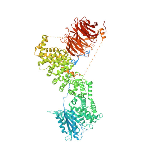
    6ZPN

  • PubMed Abstract: 

    The activation of cap-dependent translation in eukaryotes requires multisite, hierarchical phosphorylation of 4E-BP by the 1 MDa kinase mammalian target of rapamycin complex 1 (mTORC1). To resolve the mechanism of this hierarchical phosphorylation at the atomic level, we monitored by NMR spectroscopy the interaction of intrinsically disordered 4E binding protein isoform 1 (4E-BP1) with the mTORC1 subunit regulatory-associated protein of mTOR (Raptor). The N-terminal RAIP motif and the C-terminal TOR signaling (TOS) motif of 4E-BP1 bind separate sites in Raptor, resulting in avidity-based tethering of 4E-BP1. This tethering orients the flexible central region of 4E-BP1 toward the mTORC1 kinase site for phosphorylation. The structural constraints imposed by the two tethering interactions, combined with phosphorylation-induced conformational switching of 4E-BP1, explain the hierarchy of 4E-BP1 phosphorylation by mTORC1. Furthermore, we demonstrate that mTORC1 recognizes both free and eIF4E-bound 4E-BP1, allowing rapid phosphorylation of the entire 4E-BP1 pool and efficient activation of translation. Finally, our findings provide a mechanistic explanation for the differential rapamycin sensitivity of the 4E-BP1 phosphorylation sites.


  • Organizational Affiliation
    • Biozentrum, University of Basel, 4056 Basel, Switzerland.

Macromolecule Content 

  • Total Structure Weight: 339.28 kDa 
  • Atom Count: 17,406 
  • Modeled Residue Count: 2,158 
  • Deposited Residue Count: 3,008 
  • Unique protein chains: 1

Macromolecules

Find similar proteins by:|  3D Structure
Entity ID: 1
MoleculeChains  Sequence LengthOrganismDetailsImage
WD_REPEATS_REGION domain-containing protein
A, B
1,504Thermochaetoides thermophila DSM 1495Mutation(s): 0 
Gene Names: CTHT_0014610
UniProt
Find proteins for G0S1S2 (Chaetomium thermophilum (strain DSM 1495 / CBS 144.50 / IMI 039719))
Explore G0S1S2 
Go to UniProtKB:  G0S1S2
Entity Groups
Sequence Clusters30% Identity50% Identity70% Identity90% Identity95% Identity100% Identity
UniProt GroupG0S1S2
Sequence Annotations
Expand
Reference Sequence

Experimental Data & Validation

Experimental Data

  • Method: X-RAY DIFFRACTION
  • Resolution: 3.50 Å
  • R-Value Free:  0.245 (Depositor), 0.244 (DCC) 
  • R-Value Work:  0.228 (Depositor), 0.228 (DCC) 
  • R-Value Observed: 0.229 (Depositor) 
Space Group: P 41 21 2
Unit Cell:
Length ( Å )Angle ( ˚ )
a = 183.306α = 90
b = 183.306β = 90
c = 271.393γ = 90
Software Package:
Software NamePurpose
PHENIXrefinement
PDB_EXTRACTdata extraction
Cootmodel building
DIALSdata scaling
PHASERphasing
DIALSdata processing
DIALSdata reduction

Structure Validation

View Full Validation Report


Entry History 

& Funding Information

Deposition Data


Funding OrganizationLocationGrant Number
Swiss National Science FoundationSwitzerland159696
Swiss National Science FoundationSwitzerland179323

Revision History  (Full details and data files)

  • Version 1.0: 2021-05-19
    Type: Initial release
  • Version 1.1: 2021-06-16
    Changes: Database references
  • Version 1.2: 2024-01-31
    Changes: Data collection, Database references, Refinement description