6ZDV | pdb_00006zdv

Crystal structure of stabilized A2A adenosine receptor A2AR-StaR2-bRIL in complex with Chromone 5d


Experimental Data Snapshot

  • Method: X-RAY DIFFRACTION
  • Resolution: 2.13 Å
  • R-Value Free: 
    0.224 (Depositor), 0.223 (DCC) 
  • R-Value Work: 
    0.215 (Depositor), 0.220 (DCC) 
  • R-Value Observed: 
    0.215 (Depositor) 

Starting Model: experimental
View more details

wwPDB Validation 3D Report Full Report

Validation slider image for 6ZDV

Ligand Structure Quality Assessment 


This is version 1.3 of the entry. See complete history

Literature

X-Ray Crystallography and Free Energy Calculations Reveal the Binding Mechanism of A 2A Adenosine Receptor Antagonists.

Jespers, W.Verdon, G.Azuaje, J.Majellaro, M.Keranen, H.Garcia-Mera, X.Congreve, M.Deflorian, F.de Graaf, C.Zhukov, A.Dore, A.S.Mason, J.S.Aqvist, J.Cooke, R.M.Sotelo, E.Gutierrez-de-Teran, H.

(2020) Angew Chem Int Ed Engl 59: 16536-16543

  • DOI: https://doi.org/10.1002/anie.202003788
  • Primary Citation Related Structures: 
    6ZDR, 6ZDV

  • PubMed Abstract: 

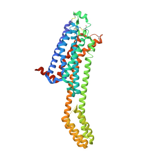
    We present a robust protocol based on iterations of free energy perturbation (FEP) calculations, chemical synthesis, biophysical mapping and X-ray crystallography to reveal the binding mode of an antagonist series to the A 2A adenosine receptor (AR). Eight A 2A AR binding site mutations from biophysical mapping experiments were initially analyzed with sidechain FEP simulations, performed on alternate binding modes. The results distinctively supported one binding mode, which was subsequently used to design new chromone derivatives. Their affinities for the A 2A AR were experimentally determined and investigated through a cycle of ligand-FEP calculations, validating the binding orientation of the different chemical substituents proposed. Subsequent X-ray crystallography of the A 2A AR with a low and a high affinity chromone derivative confirmed the predicted binding orientation. The new molecules and structures here reported were driven by free energy calculations, and provide new insights on antagonist binding to the A 2A AR, an emerging target in immuno-oncology.


  • Organizational Affiliation
    • Department of Cell and Molecular Biology, Uppsala University, BMC, Biomedical Center, Box 596, Uppsala, Sweden.

Macromolecule Content 

  • Total Structure Weight: 54.2 kDa 
  • Atom Count: 3,331 
  • Modeled Residue Count: 382 
  • Deposited Residue Count: 433 
  • Unique protein chains: 1

Macromolecules

Find similar proteins by:|  3D Structure
Entity ID: 1
MoleculeChains  Sequence LengthOrganismDetailsImage
Adenosine receptor A2a,Soluble cytochrome b562,Adenosine receptor A2a433Homo sapiensEscherichia coliMutation(s): 0 
Gene Names: ADORA2AADORA2cybC
Membrane Entity: Yes 
UniProt & NIH Common Fund Data Resources
Find proteins for P0ABE7 (Escherichia coli)
Explore P0ABE7 
Go to UniProtKB:  P0ABE7
Find proteins for P29274 (Homo sapiens)
Explore P29274 
Go to UniProtKB:  P29274
PHAROS:  P29274
GTEx:  ENSG00000128271 
Entity Groups
Sequence Clusters30% Identity50% Identity70% Identity90% Identity95% Identity100% Identity
UniProt GroupsP0ABE7P29274
Sequence Annotations
Expand
Reference Sequence

Small Molecules

Ligands 6 Unique
IDChains Name / Formula / InChI Key2D Diagram3D Interactions
CLR

Query on CLR


Download:Ideal Coordinates CCD File
D [auth A],
E [auth A],
F [auth A]
CHOLESTEROL
C27 H46 O
HVYWMOMLDIMFJA-DPAQBDIFSA-N
QGW
(Subject of Investigation/LOI)

Query on QGW


Download:Ideal Coordinates CCD File
B [auth A][2-methyl-3-(4-methyl-1,3-thiazol-2-yl)-4-oxidanylidene-6-propyl-chromen-7-yl] ethanoate
C19 H19 N O4 S
NVGMNXGIQPMJSN-UHFFFAOYSA-N
OLB

Query on OLB


Download:Ideal Coordinates CCD File
G [auth A](2S)-2,3-dihydroxypropyl (9Z)-octadec-9-enoate
C21 H40 O4
RZRNAYUHWVFMIP-QJRAZLAKSA-N
OLC

Query on OLC


Download:Ideal Coordinates CCD File
V [auth A](2R)-2,3-dihydroxypropyl (9Z)-octadec-9-enoate
C21 H40 O4
RZRNAYUHWVFMIP-GDCKJWNLSA-N
OLA

Query on OLA


Download:Ideal Coordinates CCD File
H [auth A]
I [auth A]
J [auth A]
K [auth A]
L [auth A]
H [auth A],
I [auth A],
J [auth A],
K [auth A],
L [auth A],
M [auth A],
N [auth A],
O [auth A],
P [auth A],
Q [auth A],
R [auth A],
S [auth A],
T [auth A],
U [auth A]
OLEIC ACID
C18 H34 O2
ZQPPMHVWECSIRJ-KTKRTIGZSA-N
NA

Query on NA


Download:Ideal Coordinates CCD File
C [auth A]SODIUM ION
Na
FKNQFGJONOIPTF-UHFFFAOYSA-N

Experimental Data & Validation

Experimental Data

  • Method: X-RAY DIFFRACTION
  • Resolution: 2.13 Å
  • R-Value Free:  0.224 (Depositor), 0.223 (DCC) 
  • R-Value Work:  0.215 (Depositor), 0.220 (DCC) 
  • R-Value Observed: 0.215 (Depositor) 
Space Group: C 2 2 21
Unit Cell:
Length ( Å )Angle ( ˚ )
a = 39.414α = 90
b = 179.833β = 90
c = 140.085γ = 90
Software Package:
Software NamePurpose
BUSTERrefinement
XDSdata reduction
Aimlessdata scaling
PHASERphasing

Structure Validation

View Full Validation Report


Ligand Structure Quality Assessment 


Entry History 

Revision History  (Full details and data files)

  • Version 1.0: 2020-09-16
    Type: Initial release
  • Version 1.1: 2020-12-16
    Changes: Database references
  • Version 1.2: 2024-01-24
    Changes: Data collection, Database references, Refinement description
  • Version 1.3: 2024-10-23
    Changes: Structure summary