6Z85

inhibitory human GTP cyclohydrolase I - GFRP complex


Experimental Data Snapshot

  • Method: ELECTRON MICROSCOPY
  • Resolution: 2.90 Å
  • Aggregation State: PARTICLE 
  • Reconstruction Method: SINGLE PARTICLE 

wwPDB Validation   3D Report Full Report


This is version 1.1 of the entry. See complete history


Literature

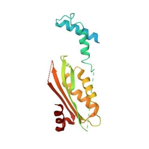
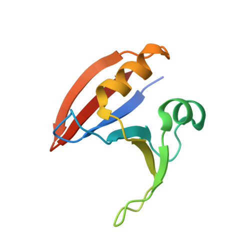
A hybrid approach reveals the allosteric regulation of GTP cyclohydrolase I.

Ebenhoch, R.Prinz, S.Kaltwasser, S.Mills, D.J.Meinecke, R.Rubbelke, M.Reinert, D.Bauer, M.Weixler, L.Zeeb, M.Vonck, J.Nar, H.

(2020) Proc Natl Acad Sci U S A 117: 31838-31849

  • DOI: https://doi.org/10.1073/pnas.2013473117
  • Primary Citation of Related Structures:  
    6Z80, 6Z85, 6Z86, 6Z87, 6Z88, 6Z89, 7ACC, 7AL9, 7ALA, 7ALB, 7ALC

  • PubMed Abstract: 

    Guanosine triphosphate (GTP) cyclohydrolase I (GCH1) catalyzes the conversion of GTP to dihydroneopterin triphosphate (H2NTP), the initiating step in the biosynthesis of tetrahydrobiopterin (BH4). Besides other roles, BH4 functions as cofactor in neurotransmitter biosynthesis. The BH4 biosynthetic pathway and GCH1 have been identified as promising targets to treat pain disorders in patients. The function of mammalian GCH1s is regulated by a metabolic sensing mechanism involving a regulator protein, GCH1 feedback regulatory protein (GFRP). GFRP binds to GCH1 to form inhibited or activated complexes dependent on availability of cofactor ligands, BH4 and phenylalanine, respectively. We determined high-resolution structures of human GCH1-GFRP complexes by cryoelectron microscopy (cryo-EM). Cryo-EM revealed structural flexibility of specific and relevant surface lining loops, which previously was not detected by X-ray crystallography due to crystal packing effects. Further, we studied allosteric regulation of isolated GCH1 by X-ray crystallography. Using the combined structural information, we are able to obtain a comprehensive picture of the mechanism of allosteric regulation. Local rearrangements in the allosteric pocket upon BH4 binding result in drastic changes in the quaternary structure of the enzyme, leading to a more compact, tense form of the inhibited protein, and translocate to the active site, leading to an open, more flexible structure of its surroundings. Inhibition of the enzymatic activity is not a result of hindrance of substrate binding, but rather a consequence of accelerated substrate binding kinetics as shown by saturation transfer difference NMR (STD-NMR) and site-directed mutagenesis. We propose a dissociation rate controlled mechanism of allosteric, noncompetitive inhibition.


  • Organizational Affiliation

    Medicinal Chemistry, Boehringer Ingelheim Pharma GmbH & Co. KG, 88397 Biberach an der Riss, Germany.


Macromolecules
Find similar proteins by:  (by identity cutoff)  |  3D Structure
Entity ID: 1
MoleculeChains Sequence LengthOrganismDetailsImage
GTP cyclohydrolase 1224Homo sapiensMutation(s): 0 
Gene Names: GCH1DYT5GCH
EC: 3.5.4.16
UniProt & NIH Common Fund Data Resources
Find proteins for P30793 (Homo sapiens)
Explore P30793 
Go to UniProtKB:  P30793
PHAROS:  P30793
GTEx:  ENSG00000131979 
Entity Groups  
Sequence Clusters30% Identity50% Identity70% Identity90% Identity95% Identity100% Identity
UniProt GroupP30793
Sequence Annotations
Expand
  • Reference Sequence
Find similar proteins by:  (by identity cutoff)  |  3D Structure
Entity ID: 2
MoleculeChains Sequence LengthOrganismDetailsImage
GTP cyclohydrolase 1 feedback regulatory protein87Homo sapiensMutation(s): 0 
Gene Names: GCHFRGFRP
UniProt & NIH Common Fund Data Resources
Find proteins for P30047 (Homo sapiens)
Explore P30047 
Go to UniProtKB:  P30047
PHAROS:  P30047
GTEx:  ENSG00000137880 
Entity Groups  
Sequence Clusters30% Identity50% Identity70% Identity90% Identity95% Identity100% Identity
UniProt GroupP30047
Sequence Annotations
Expand
  • Reference Sequence
Small Molecules
Ligands 2 Unique
IDChains Name / Formula / InChI Key2D Diagram3D Interactions
HBI (Subject of Investigation/LOI)
Query on HBI

Download Ideal Coordinates CCD File 
BA [auth B]
DA [auth E]
FA [auth G]
HA [auth H]
JA [auth I]
BA [auth B],
DA [auth E],
FA [auth G],
HA [auth H],
JA [auth I],
LA [auth J],
NA [auth F],
V [auth A],
X [auth C],
Z [auth D]
7,8-DIHYDROBIOPTERIN
C9 H13 N5 O3
FEMXZDUTFRTWPE-DZSWIPIPSA-N
ZN
Query on ZN

Download Ideal Coordinates CCD File 
AA [auth B]
CA [auth E]
EA [auth G]
GA [auth H]
IA [auth I]
AA [auth B],
CA [auth E],
EA [auth G],
GA [auth H],
IA [auth I],
KA [auth J],
MA [auth F],
U [auth A],
W [auth C],
Y [auth D]
ZINC ION
Zn
PTFCDOFLOPIGGS-UHFFFAOYSA-N
Experimental Data & Validation

Experimental Data

  • Method: ELECTRON MICROSCOPY
  • Resolution: 2.90 Å
  • Aggregation State: PARTICLE 
  • Reconstruction Method: SINGLE PARTICLE 
EM Software:
TaskSoftware PackageVersion
MODEL REFINEMENTPHENIX1.17.1
RECONSTRUCTIONRELION3.0

Structure Validation

View Full Validation Report


Entry History 

Deposition Data

Revision History  (Full details and data files)

  • Version 1.0: 2020-12-09
    Type: Initial release
  • Version 1.1: 2020-12-23
    Changes: Database references