6Z3X | pdb_00006z3x

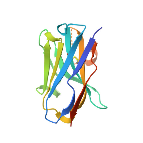
Crystal structure of the designed antibody DesAb-anti-HSA-P1


Experimental Data Snapshot

  • Method: X-RAY DIFFRACTION
  • Resolution: 1.74 Å
  • R-Value Free: 
    0.220 (Depositor), 0.228 (DCC) 
  • R-Value Work: 
    0.193 (Depositor), 0.197 (DCC) 
  • R-Value Observed: 
    0.194 (Depositor) 

Starting Model: experimental
View more details

wwPDB Validation 3D Report Full Report

Validation slider image for 6Z3X

This is version 1.3 of the entry. See complete history. 

Literature

Fragment-based computational design of antibodies targeting structured epitopes.

Aguilar Rangel, M., Bedwell, A., Costanzi, E., Taylor, R.J., Russo, R., Bernardes, G.J.L., Ricagno, S., Frydman, J., Vendruscolo, M., Sormanni, P.

(2022) Sci Adv 8: eabp9540-eabp9540

  • DOI: https://doi.org/10.1126/sciadv.abp9540
  • Primary Citation Related Structures: 
    6Z3X

  • PubMed Abstract: 

    De novo design methods hold the promise of reducing the time and cost of antibody discovery while enabling the facile and precise targeting of predetermined epitopes. Here, we describe a fragment-based method for the combinatorial design of antibody binding loops and their grafting onto antibody scaffolds. We designed and tested six single-domain antibodies targeting different epitopes on three antigens, including the receptor-binding domain of the SARS-CoV-2 spike protein. Biophysical characterization showed that all designs are stable and bind their intended targets with affinities in the nanomolar range without in vitro affinity maturation. We further discuss how a high-resolution input antigen structure is not required, as similar predictions are obtained when the input is a crystal structure or a computer-generated model. This computational procedure, which readily runs on a laptop, provides a starting point for the rapid generation of lead antibodies binding to preselected epitopes.


  • Organizational Affiliation: 
    • Centre for Misfolding Diseases, Yusuf Hamied Department of Chemistry, University of Cambridge, Cambridge CB2 1EW, UK.

Macromolecule Content 

  • Total Structure Weight: 30.98 kDa 
  • Atom Count: 2,037 
  • Modeled Residue Count: 240 
  • Deposited Residue Count: 274 
  • Unique protein chains: 1

Macromolecules

Find similar proteins by:|  3D Structure
Entity ID: 1
MoleculeChains  Sequence LengthOrganismDetailsImage
DesAb-anti-HSA-P1
A, B
137synthetic constructMutation(s): 0 
Entity Groups
Sequence Clusters30% Identity50% Identity70% Identity90% Identity95% Identity100% Identity
Sequence Annotations
Expand
Reference Sequence

Small Molecules

Ligands 3 Unique
IDChains Name / Formula / InChI Key2D Diagram3D Interactions
CAC

Query on CAC


Download:Ideal Coordinates CCD File
C [auth A]
D [auth B]
E [auth B]
F [auth B]
H [auth B]
C [auth A],
D [auth B],
E [auth B],
F [auth B],
H [auth B],
I [auth B]
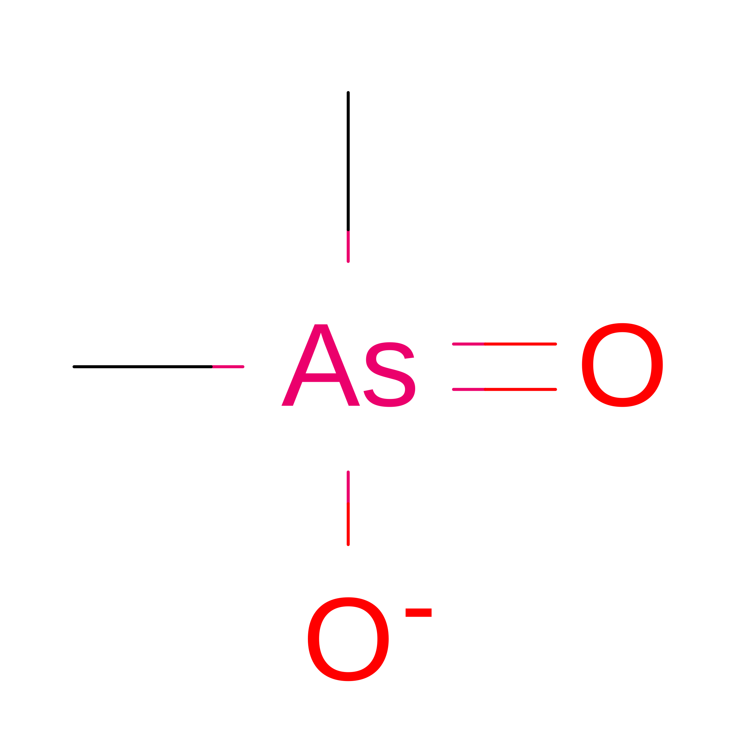
CACODYLATE ION
C2 H6 As O2
OGGXGZAMXPVRFZ-UHFFFAOYSA-M
IMD

Query on IMD


Download:Ideal Coordinates CCD File
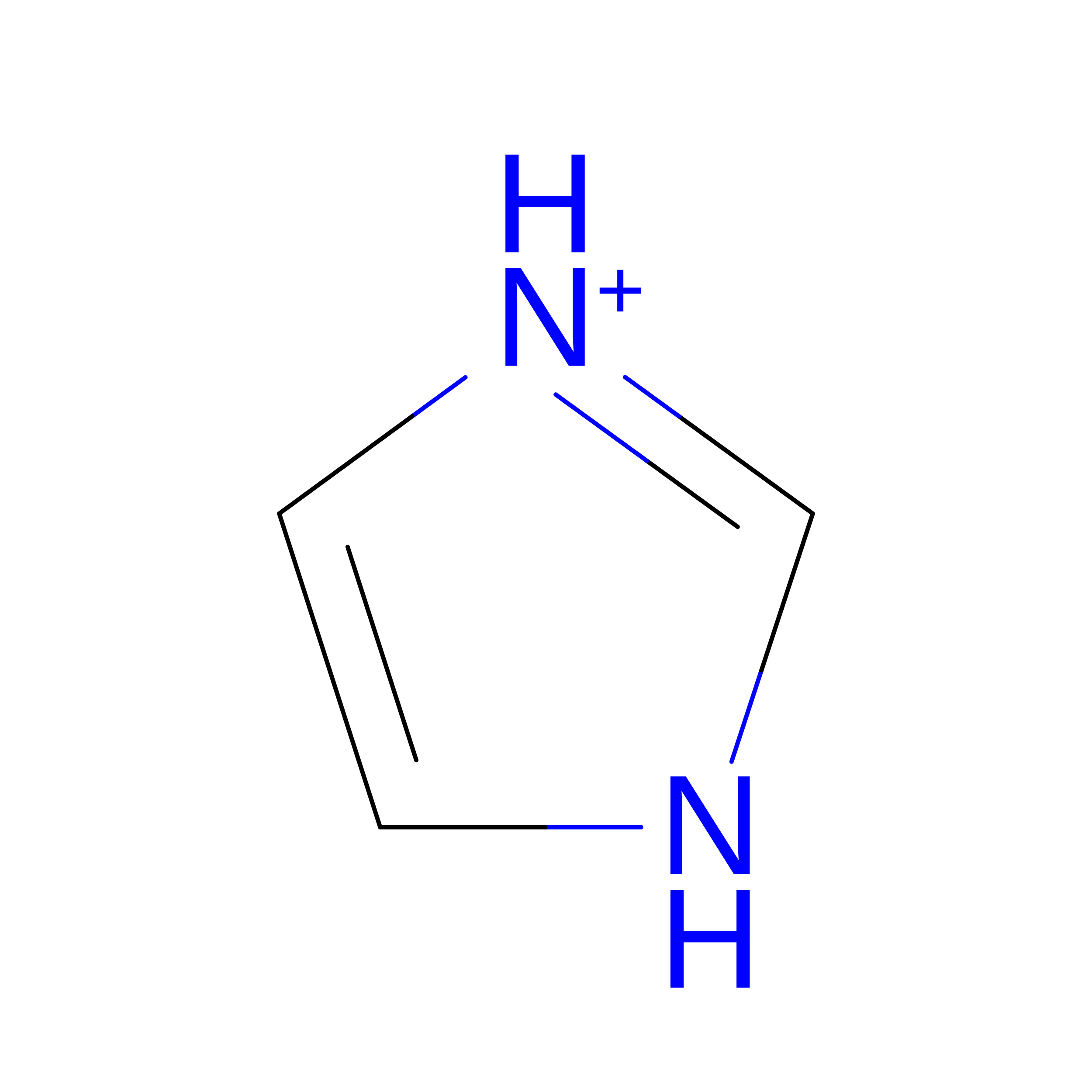
G [auth B]IMIDAZOLE
C3 H5 N2
RAXXELZNTBOGNW-UHFFFAOYSA-O
NA

Query on NA


Download:Ideal Coordinates CCD File
J [auth B]SODIUM ION
Na
FKNQFGJONOIPTF-UHFFFAOYSA-N

Experimental Data & Validation

Experimental Data

  • Method: X-RAY DIFFRACTION
  • Resolution: 1.74 Å
  • R-Value Free:  0.220 (Depositor), 0.228 (DCC) 
  • R-Value Work:  0.193 (Depositor), 0.197 (DCC) 
  • R-Value Observed: 0.194 (Depositor) 
Space Group: P 21 21 21
Unit Cell:
Length ( Å )Angle ( ˚ )
a = 40.718α = 90
b = 52.411β = 90
c = 99.176γ = 90
Software Package:
Software NamePurpose
PHENIXrefinement
PHENIXrefinement
DIALSdata reduction
Aimlessdata scaling
PHASERphasing

Structure Validation

View Full Validation Report


Entry History 

Deposition Data

Revision History  (Full details and data files)

  • Version 1.0: 2021-03-31
    Type: Initial release
  • Version 1.1: 2022-11-23
    Changes: Database references
  • Version 1.2: 2024-01-31
    Changes: Data collection, Refinement description
  • Version 1.3: 2024-11-13
    Changes: Structure summary