The Antibacterial Type VII Secretion System of Bacillus subtilis: Structure and Interactions of the Pseudokinase YukC/EssB.
Tassinari, M., Doan, T., Bellinzoni, M., Chabalier, M., Ben-Assaya, M., Martinez, M., Gaday, Q., Alzari, P.M., Cascales, E., Fronzes, R., Gubellini, F.(2022) mBio 13: e0013422-e0013422
- PubMed: 36154281 Search on PubMedSearch on PubMed Central
- DOI: https://doi.org/10.1128/mbio.00134-22
- Primary Citation Related Structures:
6Z0F - PubMed Abstract:
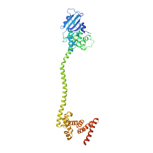
Type VIIb secretion systems (T7SSb) were recently proposed to mediate different aspects of Firmicutes physiology, including bacterial pathogenicity and competition. However, their architecture and mechanism of action remain largely obscure. Here, we present a detailed analysis of the T7SSb-mediated bacterial competition in Bacillus subtilis, using the effector YxiD as a model for the LXG secreted toxins. By systematically investigating protein-protein interactions, we reveal that the membrane subunit YukC contacts all T7SSb components, including the WXG100 substrate YukE and the LXG effector YxiD. YukC's crystal structure shows unique features, suggesting an intrinsic flexibility that is required for T7SSb antibacterial activity. Overall, our results shed light on the role and molecular organization of the T7SSb and demonstrate the potential of B. subtilis as a model system for extensive structure-function studies of these secretion machineries. IMPORTANCE Type VII secretion systems mediate protein extrusion from Gram-positive bacteria and are classified as T7SSa and T7SSb in Actinobacteria and in Firmicutes , respectively. Despite the genetic divergence of T7SSa and T7SSb, the high degree of structural similarity of their WXG100 substrates suggests similar secretion mechanisms. Recent advances revealed the structures of several T7SSa cytoplasmic membrane complexes, but the molecular mechanism of secretion and the T7SSb architecture remain obscure. Here, we provide hints on the organization of T7SSb in B. subtilis and a high-resolution structure of its central pseudokinase subunit, opening new perspectives for the understanding of the T7SSb secretion mechanism by using B. subtilis as an amenable bacterial model.
- Unité de Microbiologie Structurale, Institut Pasteurgrid.428999.7, UMR 3528, CNRS, Université de Paris, Paris, France.
Organizational Affiliation: