6YJK | pdb_00006yjk

Structure of CYRI-B (FAM49B) from Rhincodon typus


Experimental Data Snapshot

  • Method: X-RAY DIFFRACTION
  • Resolution: 2.37 Å
  • R-Value Free: 
    0.277 (Depositor), 0.277 (DCC) 
  • R-Value Work: 
    0.225 (Depositor), 0.225 (DCC) 
  • R-Value Observed: 
    0.228 (Depositor) 

Starting Model: experimental
View more details

wwPDB Validation 3D Report Full Report

Validation slider image for 6YJK

This is version 1.2 of the entry. See complete history

Literature

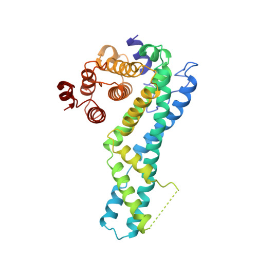
Structure of CYRI-B (FAM49B), a key regulator of cellular actin assembly.

Kaplan, E.Stone, R.Hume, P.J.Greene, N.P.Koronakis, V.

(2020) Acta Crystallogr D Struct Biol 76: 1015-1024

  • DOI: https://doi.org/10.1107/S2059798320010906
  • Primary Citation Related Structures: 
    6YJJ, 6YJK

  • PubMed Abstract: 

    In eukaryotes, numerous fundamental processes are controlled by the WAVE regulatory complex (WRC) that regulates cellular actin polymerization, crucial for cell motility, cell-cell adhesion and epithelial differentiation. Actin assembly is triggered by interaction of the small GTPase Rac1 with CYFIP1, a key component of the WRC. Previously known as FAM49B, CYRI-B is a protein that is highly conserved across the Eukaryota and has recently been revealed to be a key regulator of Rac1 activity. Mutation of CYRI-B or alteration of its expression therefore leads to altered actin nucleation dynamics, with impacts on lamellipodia formation, cell migration and infection by intracellular pathogens. In addition, knockdown of CYRI-B expression in cancer cell lines results in accelerated cell proliferation and invasiveness. Here, the structure of Rhincodon typus (whale shark) CYRI-B is presented, which is the first to be reported of any CYRI family member. Solved by X-ray crystallography, the structure reveals that CYRI-B comprises three distinct α-helical subdomains and is highly structurally related to a conserved domain present in CYFIP proteins. The work presented here establishes a template towards a better understanding of CYRI-B biological function.


  • Organizational Affiliation
    • Department of Pathology, University of Cambridge, Tennis Court Road, Cambridge CB2 1QP, United Kingdom.

Macromolecule Content 

  • Total Structure Weight: 36.91 kDa 
  • Atom Count: 2,581 
  • Modeled Residue Count: 320 
  • Deposited Residue Count: 325 
  • Unique protein chains: 1

Macromolecules

Find similar proteins by:|  3D Structure
Entity ID: 1
MoleculeChains  Sequence LengthOrganismDetailsImage
CYRI-B (FAM49B)325Rhincodon typusMutation(s): 0 
Gene Names: fam49b
UniProt
Find proteins for A0A7M4DUF3 (Rhincodon typus)
Explore A0A7M4DUF3 
Go to UniProtKB:  A0A7M4DUF3
Entity Groups
Sequence Clusters30% Identity50% Identity70% Identity90% Identity95% Identity100% Identity
UniProt GroupA0A7M4DUF3
Sequence Annotations
Expand
Reference Sequence

Experimental Data & Validation

Experimental Data

  • Method: X-RAY DIFFRACTION
  • Resolution: 2.37 Å
  • R-Value Free:  0.277 (Depositor), 0.277 (DCC) 
  • R-Value Work:  0.225 (Depositor), 0.225 (DCC) 
  • R-Value Observed: 0.228 (Depositor) 
Space Group: P 21 21 21
Unit Cell:
Length ( Å )Angle ( ˚ )
a = 39.81α = 90
b = 72.23β = 90
c = 107.81γ = 90
Software Package:
Software NamePurpose
PDB_EXTRACTdata extraction
PHENIXrefinement
REFMACrefinement
PHASERphasing
Aimlessdata scaling
iMOSFLMdata reduction

Structure Validation

View Full Validation Report


Entry History 

& Funding Information

Deposition Data

  • Released Date: 2020-10-14 
  • Deposition Author(s): Kaplan, E.

Funding OrganizationLocationGrant Number
Medical Research Council (MRC, United Kingdom)United KingdomMR/N000994/1
Wellcome TrustUnited Kingdom101828/Z/13/Z

Revision History  (Full details and data files)

  • Version 1.0: 2020-10-14
    Type: Initial release
  • Version 1.1: 2020-10-21
    Changes: Database references
  • Version 1.2: 2024-01-24
    Changes: Data collection, Database references, Refinement description