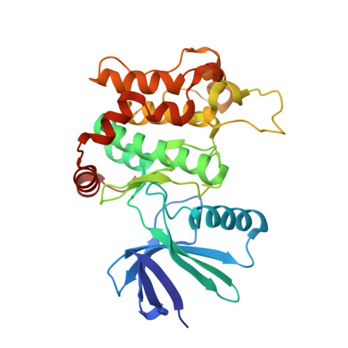
6YG2

Crystal structure of MKK7 (MAP2K7) in complex with ibrutnib, with covalent and allosteric binding modes


Experimental Data Snapshot

  • Method: X-RAY DIFFRACTION
  • Resolution: 2.00 Å
  • R-Value Free: 0.230 
  • R-Value Work: 0.179 
  • R-Value Observed: 0.181 

Starting Model: experimental
View more details

wwPDB Validation   3D Report Full Report


Ligand Structure Quality Assessment 


This is version 1.3 of the entry. See complete history


Literature

Catalytic Domain Plasticity of MKK7 Reveals Structural Mechanisms of Allosteric Activation and Diverse Targeting Opportunities.

Schroder, M.Tan, L.Wang, J.Liang, Y.Gray, N.S.Knapp, S.Chaikuad, A.

(2020) Cell Chem Biol 27: 1285-1295.e4

  • DOI: https://doi.org/10.1016/j.chembiol.2020.07.014
  • Primary Citation of Related Structures:  
    6YFZ, 6YG0, 6YG1, 6YG2, 6YG3, 6YG4, 6YG5, 6YG6, 6YG7, 6YZ4

  • PubMed Abstract: 

    MKK7 (MEK7) is a key regulator of the JNK stress signaling pathway and targeting MKK7 has been proposed as a chemotherapeutic strategy. Detailed understanding of the MKK7 structure and factors that affect its activity is therefore of critical importance. Here, we present a comprehensive set of MKK7 crystal structures revealing insights into catalytic domain plasticity and the role of the N-terminal regulatory helix, conserved in all MAP2Ks, mediating kinase activation. Crystal structures harboring this regulatory helix revealed typical structural features of active kinase, providing exclusively a first model of the MAP2K active state. A small-molecule screening campaign yielded multiple scaffolds, including type II irreversible inhibitors a binding mode that has not been reported previously. We also observed an unprecedented allosteric pocket located in the N-terminal lobe for the approved drug ibrutinib. Collectively, our structural and functional data expand and provide alternative targeting strategies for this important MAP2K kinase.


  • Organizational Affiliation

    Institute of Pharmaceutical Chemistry, Goethe-University Frankfurt, Frankfurt 60438, Germany; Structural Genomics Consortium, BMLS, Goethe-University Frankfurt, Frankfurt 60438, Germany.


Macromolecules
Find similar proteins by:  (by identity cutoff)  |  3D Structure
Entity ID: 1
MoleculeChains Sequence LengthOrganismDetailsImage
Dual specificity mitogen-activated protein kinase kinase 7307Homo sapiensMutation(s): 0 
Gene Names: MAP2K7JNKK2MEK7MKK7PRKMK7SKK4
EC: 2.7.12.2
UniProt & NIH Common Fund Data Resources
Find proteins for O14733 (Homo sapiens)
Explore O14733 
Go to UniProtKB:  O14733
PHAROS:  O14733
GTEx:  ENSG00000076984 
Entity Groups  
Sequence Clusters30% Identity50% Identity70% Identity90% Identity95% Identity100% Identity
UniProt GroupO14733
Sequence Annotations
Expand
  • Reference Sequence
Small Molecules
Ligands 4 Unique
IDChains Name / Formula / InChI Key2D Diagram3D Interactions
8E8 (Subject of Investigation/LOI)
Query on 8E8

Download Ideal Coordinates CCD File 
O [auth A]1-[(3~{R})-3-[4-azanyl-3-(4-phenoxyphenyl)pyrazolo[3,4-d]pyrimidin-1-yl]piperidin-1-yl]propan-1-one
C25 H26 N6 O2
ROGRQCNRPWIQJN-GOSISDBHSA-N
1E8 (Subject of Investigation/LOI)
Query on 1E8

Download Ideal Coordinates CCD File 
N [auth A]1-{(3R)-3-[4-amino-3-(4-phenoxyphenyl)-1H-pyrazolo[3,4-d]pyrimidin-1-yl]piperidin-1-yl}prop-2-en-1-one
C25 H24 N6 O2
XYFPWWZEPKGCCK-GOSISDBHSA-N
DMS
Query on DMS

Download Ideal Coordinates CCD File 
J [auth A],
K [auth A],
L [auth A],
M [auth A]
DIMETHYL SULFOXIDE
C2 H6 O S
IAZDPXIOMUYVGZ-UHFFFAOYSA-N
EDO
Query on EDO

Download Ideal Coordinates CCD File 
B [auth A]
C [auth A]
D [auth A]
E [auth A]
F [auth A]
B [auth A],
C [auth A],
D [auth A],
E [auth A],
F [auth A],
G [auth A],
H [auth A],
I [auth A]
1,2-ETHANEDIOL
C2 H6 O2
LYCAIKOWRPUZTN-UHFFFAOYSA-N
Experimental Data & Validation

Experimental Data

  • Method: X-RAY DIFFRACTION
  • Resolution: 2.00 Å
  • R-Value Free: 0.230 
  • R-Value Work: 0.179 
  • R-Value Observed: 0.181 
  • Space Group: P 21 21 21
Unit Cell:
Length ( Å )Angle ( ˚ )
a = 53.111α = 90
b = 74.955β = 90
c = 86.666γ = 90
Software Package:
Software NamePurpose
REFMACrefinement
SCALAdata scaling
PDB_EXTRACTdata extraction
XDSdata reduction
PHASERphasing

Structure Validation

View Full Validation Report


Ligand Structure Quality Assessment 


Entry History 

Deposition Data

Revision History  (Full details and data files)

  • Version 1.0: 2020-08-12
    Type: Initial release
  • Version 1.1: 2020-09-02
    Changes: Database references
  • Version 1.2: 2020-10-28
    Changes: Database references
  • Version 1.3: 2024-01-24
    Changes: Data collection, Database references, Refinement description