6Y90

Structure of full-length CD20 in complex with Rituximab Fab


Experimental Data Snapshot

  • Method: ELECTRON MICROSCOPY
  • Resolution: 3.69 Å
  • Aggregation State: PARTICLE 
  • Reconstruction Method: SINGLE PARTICLE 

wwPDB Validation   3D Report Full Report


This is version 1.0 of the entry. See complete history


Literature

Binding mechanisms of therapeutic antibodies to human CD20.

Kumar, A.Planchais, C.Fronzes, R.Mouquet, H.Reyes, N.

(2020) Science 369: 793-799

  • DOI: https://doi.org/10.1126/science.abb8008
  • Primary Citation of Related Structures:  
    6Y90, 6Y92, 6Y97, 6Y9A

  • PubMed Abstract: 

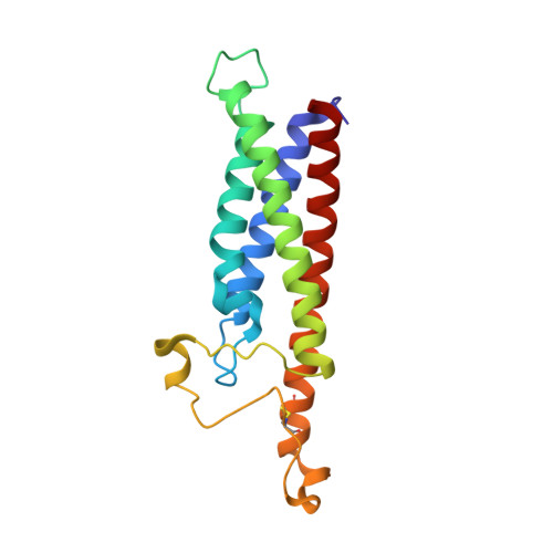
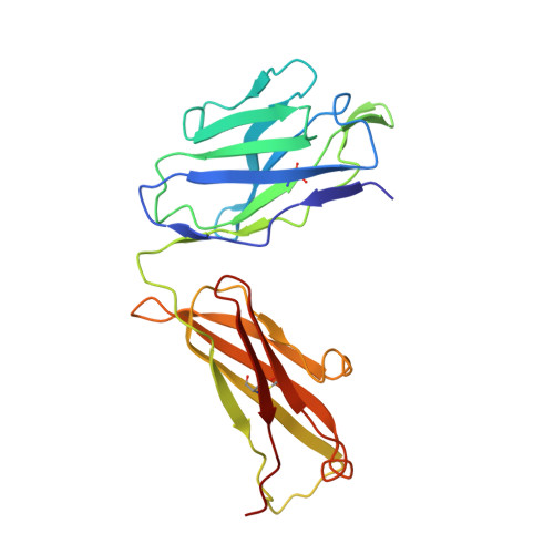
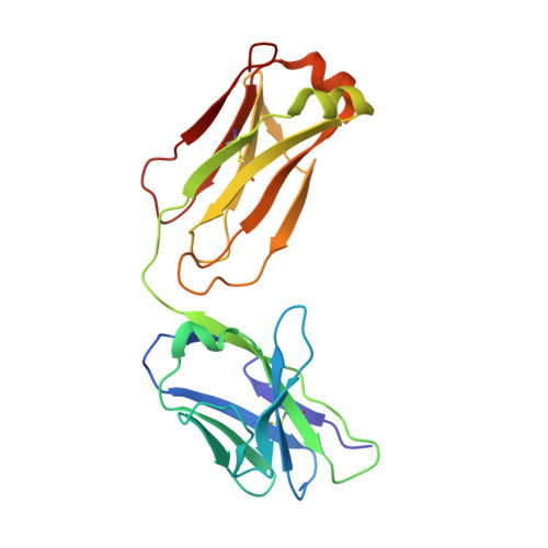
    Monoclonal antibodies (mAbs) targeting human antigen CD20 (cluster of differentiation 20) constitute important immunotherapies for the treatment of B cell malignancies and autoimmune diseases. Type I and II therapeutic mAbs differ in B cell binding properties and cytotoxic effects, reflecting differential interaction mechanisms with CD20. Here we present 3.7- to 4.7-angstrom cryo-electron microscopy structures of full-length CD20 in complexes with prototypical type I rituximab and ofatumumab and type II obinutuzumab. The structures and binding thermodynamics demonstrate that upon binding to CD20, type II mAbs form terminal complexes that preclude recruitment of additional mAbs and complement components, whereas type I complexes act as molecular seeds to increase mAb local concentration for efficient complement activation. Among type I mAbs, ofatumumab complexes display optimal geometry for complement recruitment. The uncovered mechanisms should aid rational design of next-generation immunotherapies targeting CD20.


  • Organizational Affiliation

    Membrane Protein Mechanisms Unit, Institut Pasteur, 75015 Paris, France.


Macromolecules
Find similar proteins by:  (by identity cutoff)  |  3D Structure
Entity ID: 1
MoleculeChains Sequence LengthOrganismDetailsImage
B-lymphocyte antigen CD20
A, B
172Homo sapiensMutation(s): 0 
Gene Names: MS4A1CD20
Membrane Entity: Yes 
UniProt & NIH Common Fund Data Resources
Find proteins for P11836 (Homo sapiens)
Explore P11836 
Go to UniProtKB:  P11836
PHAROS:  P11836
GTEx:  ENSG00000156738 
Entity Groups  
Sequence Clusters30% Identity50% Identity70% Identity90% Identity95% Identity100% Identity
UniProt GroupP11836
Sequence Annotations
Expand
  • Reference Sequence
Find similar proteins by:  (by identity cutoff)  |  3D Structure
Entity ID: 2
MoleculeChains Sequence LengthOrganismDetailsImage
Rituximab Fab Heavy ChainC,
E [auth H]
224Mus musculusMutation(s): 0 
Entity Groups  
Sequence Clusters30% Identity50% Identity70% Identity90% Identity95% Identity100% Identity
Sequence Annotations
Expand
  • Reference Sequence
Find similar proteins by:  (by identity cutoff)  |  3D Structure
Entity ID: 3
MoleculeChains Sequence LengthOrganismDetailsImage
Rituximab Fab Light ChainD,
F [auth L]
213Mus musculusMutation(s): 0 
Entity Groups  
Sequence Clusters30% Identity50% Identity70% Identity90% Identity95% Identity100% Identity
Sequence Annotations
Expand
  • Reference Sequence
Small Molecules
Ligands 3 Unique
IDChains Name / Formula / InChI Key2D Diagram3D Interactions
PC1
Query on PC1

Download Ideal Coordinates CCD File 
J [auth A],
U [auth B]
1,2-DIACYL-SN-GLYCERO-3-PHOSPHOCHOLINE
C44 H88 N O8 P
NRJAVPSFFCBXDT-HUESYALOSA-N
Y01
Query on Y01

Download Ideal Coordinates CCD File 
G [auth A]
H [auth A]
I [auth A]
P [auth A]
Q [auth B]
G [auth A],
H [auth A],
I [auth A],
P [auth A],
Q [auth B],
R [auth B],
S [auth B],
T [auth B]
CHOLESTEROL HEMISUCCINATE
C31 H50 O4
WLNARFZDISHUGS-MIXBDBMTSA-N
MYS
Query on MYS

Download Ideal Coordinates CCD File 
K [auth A]
L [auth A]
M [auth A]
N [auth A]
O [auth A]
K [auth A],
L [auth A],
M [auth A],
N [auth A],
O [auth A],
V [auth B],
W [auth B],
X [auth B],
Y [auth B],
Z [auth B]
PENTADECANE
C15 H32
YCOZIPAWZNQLMR-UHFFFAOYSA-N
Experimental Data & Validation

Experimental Data

  • Method: ELECTRON MICROSCOPY
  • Resolution: 3.69 Å
  • Aggregation State: PARTICLE 
  • Reconstruction Method: SINGLE PARTICLE 
EM Software:
TaskSoftware PackageVersion
MODEL REFINEMENTPHENIX1.17.1
RECONSTRUCTIONcryoSPARC2.14.2

Structure Validation

View Full Validation Report


Entry History & Funding Information

Deposition Data


Funding OrganizationLocationGrant Number
European Research Council (ERC)France309657

Revision History  (Full details and data files)

  • Version 1.0: 2020-08-26
    Type: Initial release