6XWY | pdb_00006xwy

Highly pH-resistant long stokes-shift, red fluorescent protein mCRISPRed


Experimental Data Snapshot

  • Method: X-RAY DIFFRACTION
  • Resolution: 1.75 Å
  • R-Value Free: 
    0.232 (Depositor), 0.238 (DCC) 
  • R-Value Work: 
    0.203 (Depositor), 0.214 (DCC) 
  • R-Value Observed: 
    0.205 (Depositor) 

Starting Model: experimental
View more details

wwPDB Validation 3D Report Full Report

Validation slider image for 6XWY

This is version 1.3 of the entry. See complete history

Literature

Targeted In Situ Protein Diversification and Intra-organelle Validation in Mammalian Cells.

Erdogan, M.Fabritius, A.Basquin, J.Griesbeck, O.

(2020) Cell Chem Biol 27: 610-621.e5

  • DOI: https://doi.org/10.1016/j.chembiol.2020.02.004
  • Primary Citation Related Structures: 
    6XWY

  • PubMed Abstract: 

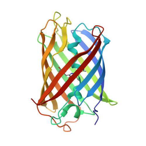
    Engineered proteins must be phenotypically selected for function in the appropriate physiological context. Here, we present a versatile approach that allows generating panels of mammalian cells that express diversified heterologous protein libraries in the cytosol or subcellular compartments under stable conditions and in a single-variant-per-cell manner. To this end we adapt CRISPR/Cas9 editing technology to diversify targeted stretches of a protein of interest in situ. We demonstrate the utility of the approach by in situ engineering and intra-lysosome specific selection of an extremely pH-resistant long Stokes shift red fluorescent protein variant. Tailoring properties to specific conditions of cellular sub-compartments or organelles of mammalian cells can be an important asset to optimize various proteins, protein-based tools, and biosensors for distinct functions.


  • Organizational Affiliation
    • Tools for Bio-Imaging, Max-Planck-Institut für Neurobiologie, Am Klopferspitz 18, Martinsried 82152, Germany.

Macromolecule Content 

  • Total Structure Weight: 99.77 kDa 
  • Atom Count: 7,559 
  • Modeled Residue Count: 869 
  • Deposited Residue Count: 876 
  • Unique protein chains: 1

Macromolecules

Find similar proteins by:|  3D Structure
Entity ID: 1
MoleculeChains  Sequence LengthOrganismDetailsImage
Red fluorescent protein eqFP611
A, B, C, D
219Entacmaea quadricolorMutation(s): 1 
UniProt
Find proteins for Q8ISF8 (Entacmaea quadricolor)
Explore Q8ISF8 
Go to UniProtKB:  Q8ISF8
Entity Groups
Sequence Clusters30% Identity50% Identity70% Identity90% Identity95% Identity100% Identity
UniProt GroupQ8ISF8
Sequence Annotations
Expand
Reference Sequence

Experimental Data & Validation

Experimental Data

  • Method: X-RAY DIFFRACTION
  • Resolution: 1.75 Å
  • R-Value Free:  0.232 (Depositor), 0.238 (DCC) 
  • R-Value Work:  0.203 (Depositor), 0.214 (DCC) 
  • R-Value Observed: 0.205 (Depositor) 
Space Group: C 2 2 21
Unit Cell:
Length ( Å )Angle ( ˚ )
a = 92.02α = 90
b = 96.861β = 90
c = 232.922γ = 90
Software Package:
Software NamePurpose
PHENIXrefinement
XDSdata processing
Aimlessdata scaling
PHASERphasing

Structure Validation

View Full Validation Report


Entry History 

Deposition Data

Revision History  (Full details and data files)

  • Version 1.0: 2020-02-05
    Type: Initial release
  • Version 1.1: 2020-03-18
    Changes: Database references
  • Version 1.2: 2020-06-03
    Changes: Database references
  • Version 1.3: 2024-01-24
    Changes: Data collection, Database references, Refinement description