6XU4 | pdb_00006xu4

Crystal structure of the genetically-encoded FGCaMP calcium indicator in its calcium-bound state


Experimental Data Snapshot

  • Method: X-RAY DIFFRACTION
  • Resolution: 3.18 Å
  • R-Value Free: 
    0.348 (Depositor), 0.343 (DCC) 
  • R-Value Work: 
    0.279 (Depositor), 0.280 (DCC) 
  • R-Value Observed: 
    0.282 (Depositor) 

Starting Model: experimental
View more details

wwPDB Validation 3D Report Full Report

Validation slider image for 6XU4

This is version 1.3 of the entry. See complete history

Literature

FGCaMP7, an Improved Version of Fungi-Based Ratiometric Calcium Indicator for In Vivo Visualization of Neuronal Activity.

Barykina, N.V.Sotskov, V.P.Gruzdeva, A.M.Wu, Y.K.Portugues, R.Subach, O.M.Chefanova, E.S.Plusnin, V.V.Ivashkina, O.I.Anokhin, K.V.Vlaskina, A.V.Korzhenevskiy, D.A.Nikolaeva, A.Y.Boyko, K.M.Rakitina, T.V.Varizhuk, A.M.Pozmogova, G.E.Subach, F.V.

(2020) Int J Mol Sci 21

  • DOI: https://doi.org/10.3390/ijms21083012
  • Primary Citation Related Structures: 
    6XU4

  • PubMed Abstract: 

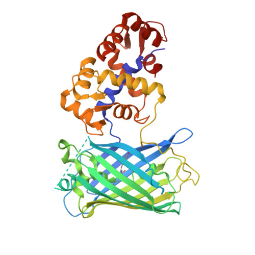
    Genetically encoded calcium indicators (GECIs) have become a widespread tool for the visualization of neuronal activity. As compared to popular GCaMP GECIs, the FGCaMP indicator benefits from calmodulin and M13-peptide from the fungi Aspergillus niger and Aspergillus fumigatus , which prevent its interaction with the intracellular environment. However, FGCaMP exhibits a two-phase fluorescence behavior with the variation of calcium ion concentration, has moderate sensitivity in neurons (as compared to the GCaMP6s indicator), and has not been fully characterized in vitro and in vivo. To address these limitations, we developed an enhanced version of FGCaMP, called FGCaMP7. FGCaMP7 preserves the ratiometric phenotype of FGCaMP, with a 3.1-fold larger ratiometric dynamic range in vitro. FGCaMP7 demonstrates 2.7- and 8.7-fold greater photostability compared to mEGFP and mTagBFP2 fluorescent proteins in vitro, respectively. The ratiometric response of FGCaMP7 is 1.6- and 1.4-fold higher, compared to the intensiometric response of GCaMP6s, in non-stimulated and stimulated neuronal cultures, respectively. We reveal the inertness of FGCaMP7 to the intracellular environment of HeLa cells using its truncated version with a deleted M13-like peptide; in contrast to the similarly truncated variant of GCaMP6s. We characterize the crystal structure of the parental FGCaMP indicator. Finally, we test the in vivo performance of FGCaMP7 in mouse brain using a two-photon microscope and an NVista miniscope; and in zebrafish using two-color ratiometric confocal imaging.


  • Organizational Affiliation
    • Laboratory for Neurobiology of Memory, P.K. Anokhin Research Institute of Normal Physiology, 125315 Moscow, Russia.

Macromolecule Content 

  • Total Structure Weight: 140.87 kDa 
  • Atom Count: 6,438 
  • Modeled Residue Count: 829 
  • Deposited Residue Count: 1,239 
  • Unique protein chains: 1

Macromolecules

Find similar proteins by:|  3D Structure
Entity ID: 1
MoleculeChains  Sequence LengthOrganismDetailsImage
FGCampA,
B,
C [auth E]
413Aspergillus nigerMutation(s): 0 
UniProt
Find proteins for P60204 (Emericella nidulans (strain FGSC A4 / ATCC 38163 / CBS 112.46 / NRRL 194 / M139))
Explore P60204 
Go to UniProtKB:  P60204
Entity Groups
Sequence Clusters30% Identity50% Identity70% Identity90% Identity95% Identity100% Identity
UniProt GroupP60204
Sequence Annotations
Expand
Reference Sequence

Small Molecules

Ligands 1 Unique
IDChains Name / Formula / InChI Key2D Diagram3D Interactions
CA
(Subject of Investigation/LOI)

Query on CA


Download:Ideal Coordinates CCD File
D [auth A]
E [auth A]
F [auth A]
G [auth A]
H [auth B]
D [auth A],
E [auth A],
F [auth A],
G [auth A],
H [auth B],
I [auth B],
J [auth B],
K [auth B],
L [auth E],
M [auth E]
CALCIUM ION
Ca
BHPQYMZQTOCNFJ-UHFFFAOYSA-N
Modified Residues  1 Unique
IDChains TypeFormula2D DiagramParent
CRO
Query on CRO
A,
B,
C [auth E]
L-PEPTIDE LINKINGC15 H17 N3 O5THR, TYR, GLY

Experimental Data & Validation

Experimental Data

  • Method: X-RAY DIFFRACTION
  • Resolution: 3.18 Å
  • R-Value Free:  0.348 (Depositor), 0.343 (DCC) 
  • R-Value Work:  0.279 (Depositor), 0.280 (DCC) 
  • R-Value Observed: 0.282 (Depositor) 
Space Group: P 43 21 2
Unit Cell:
Length ( Å )Angle ( ˚ )
a = 163.561α = 90
b = 163.561β = 90
c = 143.06γ = 90
Software Package:
Software NamePurpose
REFMACrefinement
HKL-2000data reduction
Aimlessdata scaling
MOLREPphasing
PDB_EXTRACTdata extraction

Structure Validation

View Full Validation Report


Entry History 

& Funding Information

Deposition Data


Funding OrganizationLocationGrant Number
Russian Science FoundationRussian Federation16-15-10323
Russian Foundation for Basic ResearchRussian Federation19-04-00395

Revision History  (Full details and data files)

  • Version 1.0: 2020-04-15
    Type: Initial release
  • Version 1.1: 2020-10-28
    Changes: Database references, Derived calculations
  • Version 1.2: 2024-01-24
    Changes: Data collection, Database references, Refinement description
  • Version 1.3: 2024-11-13
    Changes: Structure summary