6XSW | pdb_00006xsw

Structure of the Notch3 NRR in complex with an antibody Fab Fragment


Experimental Data Snapshot

  • Method: X-RAY DIFFRACTION
  • Resolution: 2.98 Å
  • R-Value Free: 
    0.256 (Depositor), 0.255 (DCC) 
  • R-Value Work: 
    0.233 (Depositor) 
  • R-Value Observed: 
    0.234 (Depositor) 

Starting Model: experimental
View more details

wwPDB Validation 3D Report Full Report

Validation slider image for 6XSW

Ligand Structure Quality Assessment 


This is version 1.3 of the entry. See complete history

Literature

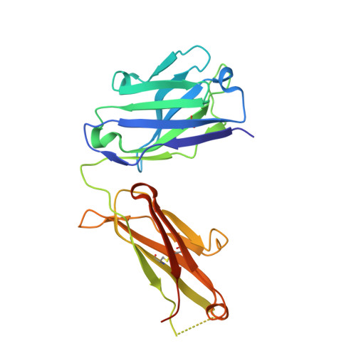
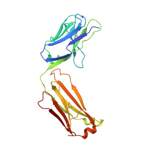
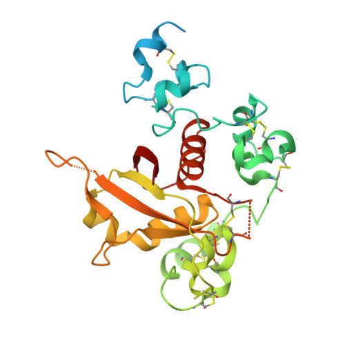
NOTCH3-targeted antibody drug conjugates regress tumors by inducing apoptosis in receptor cells and through transendocytosis into ligand cells.

Geles, K.G.Gao, Y.Giannakou, A.Sridharan, L.Yamin, T.T.Zhang, J.Karim, R.Bard, J.Piche-Nicholas, N.Charati, M.Maderna, A.Lucas, J.Golas, J.Guffroy, M.Pirie-Shepherd, S.Roy, M.Qian, J.Franks, T.Zhong, W.O'Donnell, C.J.Tchistiakova, L.Gerber, H.P.Sapra, P.

(2021) Cell Rep Med 2: 100279-100279

  • DOI: https://doi.org/10.1016/j.xcrm.2021.100279
  • Primary Citation Related Structures: 
    6XSW

  • PubMed Abstract: 

    Aberrant NOTCH3 signaling and overexpression is oncogenic, associated with cancer stem cells and drug resistance, yet therapeutic targeting remains elusive. Here, we develop NOTCH3-targeted antibody drug conjugates (NOTCH3-ADCs) by bioconjugation of an auristatin microtubule inhibitor through a protease cleavable linker to two antibodies with differential abilities to inhibit signaling. The signaling inhibitory antibody rapidly induces ligand-independent receptor clustering and internalization through both caveolin and clathrin-mediated pathways. The non-inhibitory antibody also efficiently endocytoses via clathrin without inducing receptor clustering but with slower lysosomal co-localization kinetics. In addition, DLL4 ligand binding to the NOTCH3 receptor mediates transendocytosis of NOTCH3-ADCs into ligand-expressing cells. NOTCH3-ADCs internalize into receptor and ligand cells independent of signaling and induce cell death in both cell types representing an atypical mechanism of ADC cytotoxicity. Treatment of xenografts with NOTCH3-ADCs leads to sustained tumor regressions, outperforms standard-of-care chemotherapy, and allows targeting of tumors that overexpress NOTCH3 independent of signaling inhibition.


  • Organizational Affiliation
    • Pfizer Worldwide Research and Development, Oncology Research and Development, Pearl River, NY, USA.

Macromolecule Content 

  • Total Structure Weight: 318.98 kDa 
  • Atom Count: 18,917 
  • Modeled Residue Count: 2,508 
  • Deposited Residue Count: 2,900 
  • Unique protein chains: 3

Macromolecules

Find similar proteins by:|  3D Structure
Entity ID: 1
MoleculeChains  Sequence LengthOrganismDetailsImage
Anti-N3 Fab Heavy Chain
A, D, G, H
231Rattus norvegicusMutation(s): 0 
Find similar proteins by:|  3D Structure
Entity ID: 2
MoleculeChains  Sequence LengthOrganismDetailsImage
Anti-N3 Fab Light ChainB,
E,
I,
K [auth L]
214Rattus norvegicusMutation(s): 0 
Find similar proteins by:|  3D Structure
Entity ID: 3
MoleculeChains  Sequence LengthOrganismDetailsImage
Neurogenic locus notch homolog protein 3C,
F,
J,
L [auth X]
280Homo sapiensMutation(s): 0 
Gene Names: NOTCH3
UniProt & NIH Common Fund Data Resources
Find proteins for Q9UM47 (Homo sapiens)
Explore Q9UM47 
Go to UniProtKB:  Q9UM47
PHAROS:  Q9UM47
GTEx:  ENSG00000074181 
Entity Groups
Sequence Clusters30% Identity50% Identity70% Identity90% Identity95% Identity100% Identity
UniProt GroupQ9UM47
Glycosylation
Glycosylation Sites: 1Go to GlyGen: Q9UM47-1
Sequence Annotations
Expand
Reference Sequence

Oligosaccharides

Help  
Entity ID: 4
MoleculeChains Length2D Diagram GlycosylationD Interactions
2-acetamido-2-deoxy-beta-D-glucopyranose-(1-4)-2-acetamido-2-deoxy-beta-D-glucopyranoseM [auth P]2N-Glycosylation
Glycosylation Resources
GlyTouCan: G42666HT
GlyCosmos: G42666HT
GlyGen: G42666HT

Small Molecules

Ligands 2 Unique
IDChains Name / Formula / InChI Key2D Diagram3D Interactions
NAG

Query on NAG


Download:Ideal Coordinates CCD File
Q [auth C],
U [auth F]
2-acetamido-2-deoxy-beta-D-glucopyranose
C8 H15 N O6
OVRNDRQMDRJTHS-FMDGEEDCSA-N
CA

Query on CA


Download:Ideal Coordinates CCD File
AA [auth X]
BA [auth X]
N [auth C]
O [auth C]
P [auth C]
AA [auth X],
BA [auth X],
N [auth C],
O [auth C],
P [auth C],
R [auth F],
S [auth F],
T [auth F],
V [auth J],
W [auth J],
X [auth J],
Y [auth X],
Z [auth X]
CALCIUM ION
Ca
BHPQYMZQTOCNFJ-UHFFFAOYSA-N

Experimental Data & Validation

Experimental Data

  • Method: X-RAY DIFFRACTION
  • Resolution: 2.98 Å
  • R-Value Free:  0.256 (Depositor), 0.255 (DCC) 
  • R-Value Work:  0.233 (Depositor) 
  • R-Value Observed: 0.234 (Depositor) 
Space Group: P 21 21 21
Unit Cell:
Length ( Å )Angle ( ˚ )
a = 133.652α = 90
b = 158.951β = 90
c = 159.687γ = 90
Software Package:
Software NamePurpose
BUSTERrefinement
XDSdata reduction
Aimlessdata scaling
PHASERphasing

Structure Validation

View Full Validation Report


Ligand Structure Quality Assessment 


Entry History 

Deposition Data

  • Released Date: 2021-07-21 
  • Deposition Author(s): Bard, J.

Revision History  (Full details and data files)

  • Version 1.0: 2021-07-21
    Type: Initial release
  • Version 1.1: 2023-02-01
    Changes: Database references
  • Version 1.2: 2023-10-25
    Changes: Data collection, Refinement description
  • Version 1.3: 2024-11-06
    Changes: Structure summary