


Crystal Structure of Dihydrodipicolinate synthase (DHDPS) from Brucella suis 1330
Abendroth, J., Sankaran, B., Lorimer, D.D., Horanyi, P.S., Edwards, T.E.To be published.
Experimental Data Snapshot
Starting Model: experimental
View more details
wwPDB Validation 3D Report Full Report

Entity ID: 1 | |||||
|---|---|---|---|---|---|
| Molecule | Chains | Sequence Length | Organism | Details | Image |
| 4-hydroxy-tetrahydrodipicolinate synthase | 314 | Brucella suis 1330 | Mutation(s): 0 Gene Names: dapA, BR0646, BS1330_I0642 EC: 4.3.3.7 | 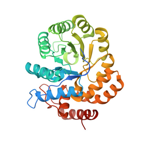 | |
UniProt | |||||
Entity Groups | |||||
| Sequence Clusters | 30% Identity50% Identity70% Identity90% Identity95% Identity100% Identity | ||||
| UniProt Group | Q8G1R0 | ||||
Sequence AnnotationsExpand | |||||
Reference Sequence | |||||
| Ligands 2 Unique | |||||
|---|---|---|---|---|---|
| ID | Chains | Name / Formula / InChI Key | 2D Diagram | 3D Interactions | |
| CIT Download:Ideal Coordinates CCD File | E [auth A], H [auth B], J [auth C], M [auth D] | CITRIC ACID C6 H8 O7 KRKNYBCHXYNGOX-UHFFFAOYSA-N |  | ||
| PO4 Download:Ideal Coordinates CCD File | F [auth A], G [auth B], I [auth B], K [auth C], L [auth D] | PHOSPHATE ION O4 P NBIIXXVUZAFLBC-UHFFFAOYSA-K |  | ||
| Length ( Å ) | Angle ( ˚ ) |
|---|---|
| a = 86.65 | α = 90 |
| b = 86.65 | β = 90 |
| c = 142.97 | γ = 120 |
| Software Name | Purpose |
|---|---|
| XDS | data reduction |
| XSCALE | data scaling |
| REFMAC | refinement |
| PDB_EXTRACT | data extraction |
| MoRDa | phasing |
| Coot | model building |


