6XCM | pdb_00006xcm

Structure of the SARS-CoV-2 spike glycoprotein in complex with the C105 neutralizing antibody Fab fragment (state 1)


Experimental Data Snapshot

  • Method: ELECTRON MICROSCOPY
  • Resolution: 3.42 Å
  • Aggregation State: PARTICLE 
  • Reconstruction Method: SINGLE PARTICLE 

Starting Model: experimental
View more details

wwPDB Validation 3D Report Full Report

Validation slider image for 6XCM

This is version 2.2 of the entry. See complete history

Literature

Structures of Human Antibodies Bound to SARS-CoV-2 Spike Reveal Common Epitopes and Recurrent Features of Antibodies.

Barnes, C.O.West Jr., A.P.Huey-Tubman, K.E.Hoffmann, M.A.G.Sharaf, N.G.Hoffman, P.R.Koranda, N.Gristick, H.B.Gaebler, C.Muecksch, F.Lorenzi, J.C.C.Finkin, S.Hagglof, T.Hurley, A.Millard, K.G.Weisblum, Y.Schmidt, F.Hatziioannou, T.Bieniasz, P.D.Caskey, M.Robbiani, D.F.Nussenzweig, M.C.Bjorkman, P.J.

(2020) Cell 182: 828

  • DOI: https://doi.org/10.1016/j.cell.2020.06.025
  • Primary Citation Related Structures: 
    6XCA, 6XCM, 6XCN

  • PubMed Abstract: 

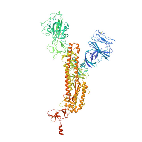
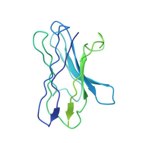
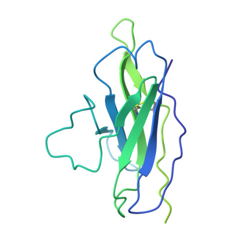
    Neutralizing antibody responses to coronaviruses mainly target the receptor-binding domain (RBD) of the trimeric spike. Here, we characterized polyclonal immunoglobulin Gs (IgGs) and Fabs from COVID-19 convalescent individuals for recognition of coronavirus spikes. Plasma IgGs differed in their focus on RBD epitopes, recognition of alpha- and beta-coronaviruses, and contributions of avidity to increased binding/neutralization of IgGs over Fabs. Using electron microscopy, we examined specificities of polyclonal plasma Fabs, revealing recognition of both S1 A and RBD epitopes on SARS-CoV-2 spike. Moreover, a 3.4 Å cryo-electron microscopy (cryo-EM) structure of a neutralizing monoclonal Fab-spike complex revealed an epitope that blocks ACE2 receptor binding. Modeling based on these structures suggested different potentials for inter-spike crosslinking by IgGs on viruses, and characterized IgGs would not be affected by identified SARS-CoV-2 spike mutations. Overall, our studies structurally define a recurrent anti-SARS-CoV-2 antibody class derived from VH3-53/VH3-66 and similarity to a SARS-CoV VH3-30 antibody, providing criteria for evaluating vaccine-elicited antibodies.


  • Organizational Affiliation
    • Division of Biology and Biological Engineering, California Institute of Technology, Pasadena, CA, USA.

Macromolecule Content 

  • Total Structure Weight: 527.03 kDa 
  • Atom Count: 26,772 
  • Modeled Residue Count: 3,390 
  • Deposited Residue Count: 4,671 
  • Unique protein chains: 3

Macromolecules

Find similar proteins by:|  3D Structure
Entity ID: 1
MoleculeChains  Sequence LengthOrganismDetailsImage
Spike glycoprotein
A, B, C
1,259Severe acute respiratory syndrome coronavirus 2Mutation(s): 0 
Gene Names: S2
UniProt
Find proteins for P0DTC2 (Severe acute respiratory syndrome coronavirus 2)
Explore P0DTC2 
Go to UniProtKB:  P0DTC2
Entity Groups
Sequence Clusters30% Identity50% Identity70% Identity90% Identity95% Identity100% Identity
UniProt GroupP0DTC2
Glycosylation
Glycosylation Sites: 14Go to GlyGen: P0DTC2-1
Sequence Annotations
Expand
Reference Sequence
Find similar proteins by:|  3D Structure
Entity ID: 2
MoleculeChains  Sequence LengthOrganismDetailsImage
C105 Fab Heavy ChainD [auth H],
E [auth N]
230Homo sapiensMutation(s): 0 
Find similar proteins by:|  3D Structure
Entity ID: 3
MoleculeChains  Sequence LengthOrganismDetailsImage
C105 Fab Light ChainF [auth L],
G [auth S]
217Homo sapiensMutation(s): 0 

Oligosaccharides

Help  
Entity ID: 4
MoleculeChains Length2D Diagram GlycosylationD Interactions
2-acetamido-2-deoxy-beta-D-glucopyranose-(1-4)-2-acetamido-2-deoxy-beta-D-glucopyranose
H [auth D],
I [auth E],
K [auth G],
L [auth I],
N [auth K],
H [auth D],
I [auth E],
K [auth G],
L [auth I],
N [auth K],
O [auth M],
P [auth O],
Q [auth P],
S [auth R]
2N-Glycosylation
Glycosylation Resources
GlyTouCan: G42666HT
GlyCosmos: G42666HT
GlyGen: G42666HT
Entity ID: 5
MoleculeChains Length2D Diagram GlycosylationD Interactions
alpha-L-fucopyranose-(1-6)-2-acetamido-2-deoxy-beta-D-glucopyranoseJ [auth F]2N-Glycosylation
Glycosylation Resources
GlyTouCan: G86851RC
GlyCosmos: G86851RC
GlyGen: G86851RC
Entity ID: 6
MoleculeChains Length2D Diagram GlycosylationD Interactions
2-acetamido-2-deoxy-beta-D-glucopyranose-(1-4)-[alpha-L-fucopyranose-(1-6)]2-acetamido-2-deoxy-beta-D-glucopyranoseM [auth J],
T
3N-Glycosylation
Glycosylation Resources
GlyTouCan: G21290RB
GlyCosmos: G21290RB
GlyGen: G21290RB
Entity ID: 7
MoleculeChains Length2D Diagram GlycosylationD Interactions
beta-D-mannopyranose-(1-4)-2-acetamido-2-deoxy-beta-D-glucopyranose-(1-4)-2-acetamido-2-deoxy-beta-D-glucopyranoseR [auth Q]3N-Glycosylation
Glycosylation Resources
GlyTouCan: G15407YE
GlyCosmos: G15407YE
GlyGen: G15407YE

Small Molecules

Ligands 1 Unique
IDChains Name / Formula / InChI Key2D Diagram3D Interactions
NAG

Query on NAG


Download:Ideal Coordinates CCD File
AA [auth A]
BA [auth A]
CA [auth A]
DA [auth A]
EA [auth B]
AA [auth A],
BA [auth A],
CA [auth A],
DA [auth A],
EA [auth B],
FA [auth B],
GA [auth B],
HA [auth B],
IA [auth B],
JA [auth B],
KA [auth B],
LA [auth B],
MA [auth B],
NA [auth B],
OA [auth B],
PA [auth C],
QA [auth C],
RA [auth C],
SA [auth C],
TA [auth C],
U [auth A],
UA [auth C],
V [auth A],
VA [auth C],
W [auth A],
WA [auth C],
X [auth A],
XA [auth C],
Y [auth A],
Z [auth A]
2-acetamido-2-deoxy-beta-D-glucopyranose
C8 H15 N O6
OVRNDRQMDRJTHS-FMDGEEDCSA-N

Experimental Data & Validation

Experimental Data

  • Method: ELECTRON MICROSCOPY
  • Resolution: 3.42 Å
  • Aggregation State: PARTICLE 
  • Reconstruction Method: SINGLE PARTICLE 
EM Software:
TaskSoftware PackageVersion
RECONSTRUCTIONcryoSPARC2.14.2
MODEL REFINEMENTPHENIX1.17

Structure Validation

View Full Validation Report


Entry History 

& Funding Information

Deposition Data


Funding OrganizationLocationGrant Number
National Institutes of Health/National Institute Of Allergy and Infectious Diseases (NIH/NIAID)United StatesP01-AI138398-S1

Revision History  (Full details and data files)

  • Version 1.0: 2020-07-01
    Type: Initial release
  • Version 1.1: 2020-07-22
    Changes: Database references
  • Version 2.0: 2020-07-29
    Type: Remediation
    Reason: Carbohydrate remediation
    Changes: Advisory, Atomic model, Data collection, Derived calculations, Structure summary
  • Version 2.1: 2020-09-02
    Changes: Database references, Structure summary
  • Version 2.2: 2024-10-16
    Changes: Data collection, Database references, Refinement description, Structure summary