Unraveling the folding and dimerization properties of the human FoxP subfamily of transcription factors.
Villalobos, P., Carvajal, A.I., Castro-Fernandez, V., Babul, J., Ramirez-Sarmiento, C.A., Medina, E.(2023) FEBS Lett 597: 1894-1905
- PubMed: 37199668 Search on PubMed
- DOI: https://doi.org/10.1002/1873-3468.14665
- Primary Citation Related Structures:
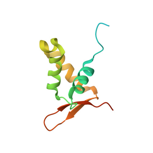
6XAT - PubMed Abstract:
Human FoxP proteins share a highly conserved DNA-binding domain that dimerizes via three-dimensional domain swapping, although showing varying oligomerization propensities among its members. Here, we present an experimental and computational characterization of all human FoxP proteins to unravel how their amino acid substitutions impact their folding and dimerization mechanism. We solved the crystal structure of the forkhead domain of FoxP4 to then perform a comparison across all members, finding that their sequence changes impact not only the structural heterogeneity of their forkhead domains but also the protein-protein association energy barrier. Lastly, we demonstrate that the accumulation of a monomeric intermediate is an oligomerization-dependent feature rather than a common aspect of monomers and dimers in this protein subfamily.
- Departamento de Biología, Facultad de Ciencias, Universidad de Chile, Santiago, Chile.
Organizational Affiliation: