6XA2

Structure of the tirandamycin C-bound P450 monooxygenase TamI


Experimental Data Snapshot

  • Method: X-RAY DIFFRACTION
  • Resolution: 2.64 Å
  • R-Value Free: 0.299 
  • R-Value Work: 0.252 
  • R-Value Observed: 0.253 

wwPDB Validation   3D Report Full Report


Ligand Structure Quality Assessment 


This is version 1.2 of the entry. See complete history


Literature

Molecular Basis of Iterative C─H Oxidation by TamI, a Multifunctional P450 monooxygenase from the Tirandamycin Biosynthetic Pathway.

Newmister, S.A.Srivastava, K.R.Espinoza, R.V.Haatveit, K.C.Khatri, Y.Martini, R.M.Garcia-Borras, M.Podust, L.M.Houk, K.N.Sherman, D.H.

(2020) ACS Catal 10: 13445-13454

  • DOI: https://doi.org/10.1021/acscatal.0c03248
  • Primary Citation of Related Structures:  
    6XA2, 6XA3

  • PubMed Abstract: 

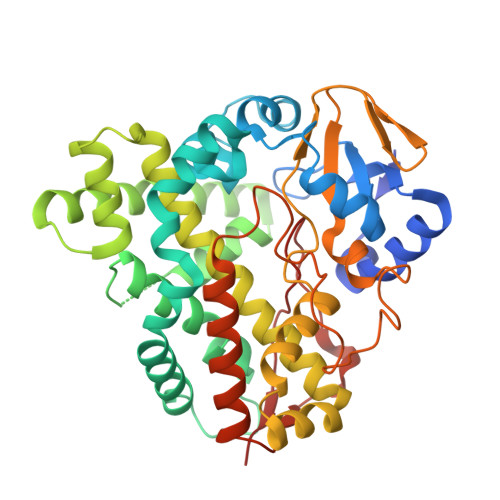
    Biocatalysis offers an expanding and powerful strategy to construct and diversify complex molecules by C─H bond functionalization. Due to their high selectivity, enzymes have become an essential tool for C─H bond functionalization and offer complementary reactivity to small-molecule catalysts. Hemoproteins, particularly cytochromes P450, have proven effective for selective oxidation of unactivated C─H bonds. Previously, we reported the in vitro characterization of an oxidative tailoring cascade in which TamI, a multifunctional P450 functions co-dependently with the TamL flavoprotein to catalyze regio- and stereoselective hydroxylations and epoxidation to yield tirandamycin A and tirandamycin B. TamI follows a defined order including 1) C10 hydroxylation, 2) C11/C12 epoxidation, and 3) C18 hydroxylation. Here we present a structural, biochemical, and computational investigation of TamI to understand the molecular basis of its substrate binding, diverse reactivity, and specific reaction sequence. The crystal structure of TamI in complex with tirandamycin C together with molecular dynamics simulations and targeted mutagenesis suggest that hydrophobic interactions with the polyene chain of its natural substrate are critical for molecular recognition. QM calculations and molecular dynamics simulations of TamI with variant substrates provided detailed information on the molecular basis of sequential reactivity, and pattern of regio- and stereo-selectivity in catalyzing the three-step oxidative cascade.


  • Organizational Affiliation

    Life Sciences Institute, University of Michigan, Ann Arbor, MI 48109, USA.


Macromolecules
Find similar proteins by:  (by identity cutoff)  |  3D Structure
Entity ID: 1
MoleculeChains Sequence LengthOrganismDetailsImage
TamI
A, B, C, D, E
A, B, C, D, E, F, G, H
414Streptomyces sp. 307-9Mutation(s): 0 
UniProt
Find proteins for D3Y1J3 (Streptomyces sp. 307-9)
Explore D3Y1J3 
Go to UniProtKB:  D3Y1J3
Entity Groups  
Sequence Clusters30% Identity50% Identity70% Identity90% Identity95% Identity100% Identity
UniProt GroupD3Y1J3
Sequence Annotations
Expand
  • Reference Sequence
Small Molecules
Ligands 2 Unique
IDChains Name / Formula / InChI Key2D Diagram3D Interactions
HEM (Subject of Investigation/LOI)
Query on HEM

Download Ideal Coordinates CCD File 
I [auth A]
K [auth B]
M [auth C]
O [auth D]
Q [auth E]
I [auth A],
K [auth B],
M [auth C],
O [auth D],
Q [auth E],
S [auth F],
U [auth G],
W [auth H]
PROTOPORPHYRIN IX CONTAINING FE
C34 H32 Fe N4 O4
KABFMIBPWCXCRK-RGGAHWMASA-L
V0A (Subject of Investigation/LOI)
Query on V0A

Download Ideal Coordinates CCD File 
J [auth A]
L [auth B]
N [auth C]
P [auth D]
R [auth E]
J [auth A],
L [auth B],
N [auth C],
P [auth D],
R [auth E],
T [auth F],
V [auth G],
X [auth H]
(3E)-3-{(2E,4E,6R)-1-hydroxy-4-methyl-6-[(1R,3R,4S,5R)-1,4,8-trimethyl-2,9-dioxabicyclo[3.3.1]non-7-en-3-yl]hepta-2,4-dien-1-ylidene}-2H-pyrrole-2,4(3H)-dione
C22 H27 N O5
MHIKRZGCDBIVFG-IVGQTCGHSA-N
Experimental Data & Validation

Experimental Data

  • Method: X-RAY DIFFRACTION
  • Resolution: 2.64 Å
  • R-Value Free: 0.299 
  • R-Value Work: 0.252 
  • R-Value Observed: 0.253 
  • Space Group: C 1 2 1
Unit Cell:
Length ( Å )Angle ( ˚ )
a = 224.554α = 90
b = 57.156β = 90.89
c = 282.659γ = 90
Software Package:
Software NamePurpose
PHENIXrefinement
PDB_EXTRACTdata extraction
Cootmodel building
XDSdata scaling
JBluIce-EPICSdata collection
XDSdata reduction
PHASERphasing

Structure Validation

View Full Validation Report


Ligand Structure Quality Assessment 


Entry History & Funding Information

Deposition Data


Funding OrganizationLocationGrant Number
National Science Foundation (NSF, United States)United StatesCHE-1700982
National Institutes of Health/National Institute of General Medical Sciences (NIH/NIGMS)United StatesR35-GM118101

Revision History  (Full details and data files)

  • Version 1.0: 2021-06-09
    Type: Initial release
  • Version 1.1: 2021-12-22
    Changes: Database references
  • Version 1.2: 2024-04-03
    Changes: Data collection, Database references, Refinement description