Structural and Functional Investigation of the Periplasmic Arsenate-Binding Protein ArrX from Chrysiogenes arsenatis .
Poddar, N., Badilla, C., Maghool, S., Osborne, T.H., Santini, J.M., Maher, M.J.(2021) Biochemistry 60: 465-476
- PubMed: 33538578 Search on PubMed
- DOI: https://doi.org/10.1021/acs.biochem.0c00555
- Primary Citation Related Structures:
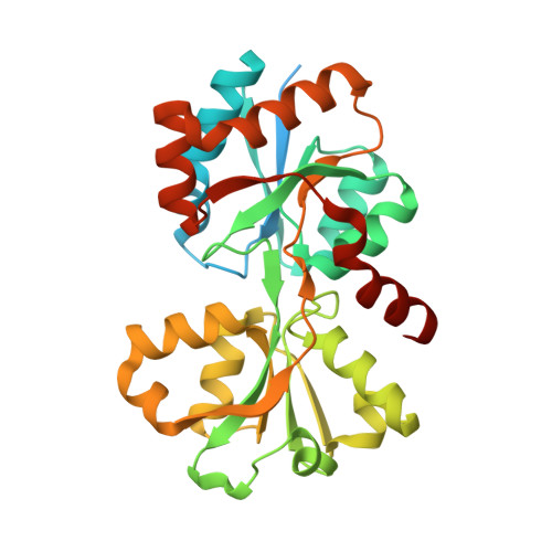
6X6B, 6X8W, 6X9G, 6XAB, 6XAD, 6XL2, 7L22 - PubMed Abstract:
The anaerobic bacterium Chrysiogenes arsenatis respires using the oxyanion arsenate (AsO 4 3- ) as the terminal electron acceptor, where it is reduced to arsenite (AsO 3 3- ) while concomitantly oxidizing various organic (e.g., acetate) electron donors. This respiratory activity is catalyzed in the periplasm of the bacterium by the enzyme arsenate reductase (Arr), with expression of the enzyme controlled by a sensor histidine kinase (ArrS) and a periplasmic-binding protein (PBP), ArrX. Here, we report for the first time, the molecular structure of ArrX in the absence and presence of bound ligand arsenate. Comparison of the ligand-bound structure of ArrX with other PBPs shows a high level of conservation of critical residues for ligand binding by these proteins; however, this suite of PBPs shows different structural alterations upon ligand binding. For ArrX and its homologue AioX (from Rhizobium sp. str. NT-26), which specifically binds arsenite, the structures of the substrate-binding sites in the vicinity of a conserved and critical cysteine residue contribute to the discrimination of binding for these chemically similar ligands.
- School of Chemistry and The Bio21 Molecular Science and Biotechnology Institute, The University of Melbourne, Parkville 3052, Australia.
Organizational Affiliation: