6X5S

Human Alpha-1,6-fucosyltransferase (FUT8) bound to GDP and A3'-Asn


Experimental Data Snapshot

  • Method: X-RAY DIFFRACTION
  • Resolution: 3.30 Å
  • R-Value Free: 0.224 
  • R-Value Work: 0.198 
  • R-Value Observed: 0.199 

wwPDB Validation   3D Report Full Report


Ligand Structure Quality Assessment 


This is version 1.3 of the entry. See complete history


Literature

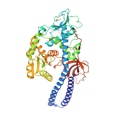
Characterizing human alpha-1,6-fucosyltransferase (FUT8) substrate specificity and structural similarities with related fucosyltransferases.

Boruah, B.M.Kadirvelraj, R.Liu, L.Ramiah, A.Li, C.Zong, G.Bosman, G.P.Yang, J.Y.Wang, L.X.Boons, G.J.Wood, Z.A.Moremen, K.W.

(2020) J Biol Chem 295: 17027-17045

  • DOI: https://doi.org/10.1074/jbc.RA120.014625
  • Primary Citation of Related Structures:  
    6X5H, 6X5R, 6X5S, 6X5T, 6X5U

  • PubMed Abstract: 

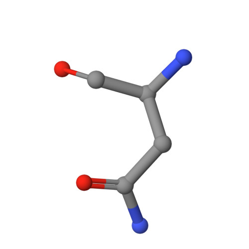
    Mammalian Asn-linked glycans are extensively processed as they transit the secretory pathway to generate diverse glycans on cell surface and secreted glycoproteins. Additional modification of the glycan core by α-1,6-fucose addition to the innermost GlcNAc residue (core fucosylation) is catalyzed by an α-1,6-fucosyltransferase (FUT8). The importance of core fucosylation can be seen in the complex pathological phenotypes of FUT8 null mice, which display defects in cellular signaling, development, and subsequent neonatal lethality. Elevated core fucosylation has also been identified in several human cancers. However, the structural basis for FUT8 substrate specificity remains unknown.Here, using various crystal structures of FUT8 in complex with a donor substrate analog, and with four distinct glycan acceptors, we identify the molecular basis for FUT8 specificity and activity. The ordering of three active site loops corresponds to an increased occupancy for bound GDP, suggesting an induced-fit folding of the donor-binding subsite. Structures of the various acceptor complexes were compared with kinetic data on FUT8 active site mutants and with specificity data from a library of glycan acceptors to reveal how binding site complementarity and steric hindrance can tune substrate affinity. The FUT8 structure was also compared with other known fucosyltransferases to identify conserved and divergent structural features for donor and acceptor recognition and catalysis. These data provide insights into the evolution of modular templates for donor and acceptor recognition among GT-B fold glycosyltransferases in the synthesis of diverse glycan structures in biological systems.


  • Organizational Affiliation

    Complex Carbohydrate Research Center, University of Georgia, Athens, Georgia, USA.


Macromolecules
Find similar proteins by:  (by identity cutoff)  |  3D Structure
Entity ID: 1
MoleculeChains Sequence LengthOrganismDetailsImage
Alpha-(1,6)-fucosyltransferase
A, B
466Homo sapiensMutation(s): 0 
Gene Names: FUT8
EC: 2.4.1.68
UniProt & NIH Common Fund Data Resources
Find proteins for Q9BYC5 (Homo sapiens)
Explore Q9BYC5 
Go to UniProtKB:  Q9BYC5
PHAROS:  Q9BYC5
GTEx:  ENSG00000033170 
Entity Groups  
Sequence Clusters30% Identity50% Identity70% Identity90% Identity95% Identity100% Identity
UniProt GroupQ9BYC5
Sequence Annotations
Expand
  • Reference Sequence

Find similar proteins by:  Sequence   |   3D Structure  

Entity ID: 2
MoleculeChains Sequence LengthOrganismDetailsImage
A3'-Asn
C, D
3Gallus gallusMutation(s): 0 
Sequence Annotations
Expand
  • Reference Sequence
Oligosaccharides

Help

Entity ID: 3
MoleculeChains Length2D Diagram Glycosylation3D Interactions
2-acetamido-2-deoxy-beta-D-glucopyranose-(1-2)-[2-acetamido-2-deoxy-beta-D-glucopyranose-(1-4)]alpha-D-mannopyranose-(1-3)-[2-acetamido-2-deoxy-beta-D-glucopyranose-(1-2)-alpha-D-mannopyranose-(1-6)]beta-D-mannopyranose-(1-4)-2-acetamido-2-deoxy-beta-D-glucopyranose-(1-4)-2-acetamido-2-deoxy-beta-D-glucopyranose
E, F
8N-Glycosylation
Glycosylation Resources
GlyTouCan:  G29011JC
GlyCosmos:  G29011JC
GlyGen:  G29011JC
Small Molecules
Ligands 3 Unique
IDChains Name / Formula / InChI Key2D Diagram3D Interactions
GDP (Subject of Investigation/LOI)
Query on GDP

Download Ideal Coordinates CCD File 
U [auth C],
V [auth D]
GUANOSINE-5'-DIPHOSPHATE
C10 H15 N5 O11 P2
QGWNDRXFNXRZMB-UUOKFMHZSA-N
GOL
Query on GOL

Download Ideal Coordinates CCD File 
T [auth B]GLYCEROL
C3 H8 O3
PEDCQBHIVMGVHV-UHFFFAOYSA-N
CL
Query on CL

Download Ideal Coordinates CCD File 
G [auth A]
H [auth A]
I [auth A]
J [auth A]
K [auth A]
G [auth A],
H [auth A],
I [auth A],
J [auth A],
K [auth A],
L [auth A],
M [auth A],
N [auth B],
O [auth B],
P [auth B],
Q [auth B],
R [auth B],
S [auth B]
CHLORIDE ION
Cl
VEXZGXHMUGYJMC-UHFFFAOYSA-M
Experimental Data & Validation

Experimental Data

  • Method: X-RAY DIFFRACTION
  • Resolution: 3.30 Å
  • R-Value Free: 0.224 
  • R-Value Work: 0.198 
  • R-Value Observed: 0.199 
  • Space Group: P 65 2 2
Unit Cell:
Length ( Å )Angle ( ˚ )
a = 150.5α = 90
b = 150.5β = 90
c = 480.31γ = 120
Software Package:
Software NamePurpose
PHENIXrefinement
XDSdata reduction
XSCALEdata scaling
PHASERphasing

Structure Validation

View Full Validation Report


Ligand Structure Quality Assessment 


Entry History & Funding Information

Deposition Data


Funding OrganizationLocationGrant Number
National Institutes of Health/National Institute on Alcohol Abuse and Alcoholism (NIH/NIAAA)United StatesR01GM130915, P41GM103390 and P01GM107012

Revision History  (Full details and data files)

  • Version 1.0: 2020-10-07
    Type: Initial release
  • Version 1.1: 2020-10-21
    Changes: Database references
  • Version 1.2: 2020-12-23
    Changes: Database references
  • Version 1.3: 2023-10-18
    Changes: Data collection, Database references, Refinement description