6WVH

Human VKOR with Brodifacoum


Experimental Data Snapshot

  • Method: X-RAY DIFFRACTION
  • Resolution: 1.99 Å
  • R-Value Free: 0.215 
  • R-Value Work: 0.195 
  • R-Value Observed: 0.196 

wwPDB Validation   3D Report Full Report


Ligand Structure Quality Assessment 


This is version 1.4 of the entry. See complete history


Literature

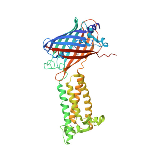
Structural basis of antagonizing the vitamin K catalytic cycle for anticoagulation.

Liu, S.Li, S.Shen, G.Sukumar, N.Krezel, A.M.Li, W.

(2021) Science 371

  • DOI: https://doi.org/10.1126/science.abc5667
  • Primary Citation of Related Structures:  
    6WV3, 6WV4, 6WV5, 6WV6, 6WV7, 6WV8, 6WV9, 6WVA, 6WVB, 6WVH, 6WVI

  • PubMed Abstract: 

    Vitamin K antagonists are widely used anticoagulants that target vitamin K epoxide reductases (VKOR), a family of integral membrane enzymes. To elucidate their catalytic cycle and inhibitory mechanism, we report 11 x-ray crystal structures of human VKOR and pufferfish VKOR-like, with substrates and antagonists in different redox states. Substrates entering the active site in a partially oxidized state form cysteine adducts that induce an open-to-closed conformational change, triggering reduction. Binding and catalysis are facilitated by hydrogen-bonding interactions in a hydrophobic pocket. The antagonists bind specifically to the same hydrogen-bonding residues and induce a similar closed conformation. Thus, vitamin K antagonists act through mimicking the key interactions and conformational changes required for the VKOR catalytic cycle.


  • Organizational Affiliation

    Department of Biochemistry and Molecular Biophysics, Washington University School of Medicine, St. Louis, MO 63110, USA.


Macromolecules
Find similar proteins by:  (by identity cutoff)  |  3D Structure
Entity ID: 1
MoleculeChains Sequence LengthOrganismDetailsImage
Vitamin K epoxide reductase, termini restrained by green fluorescent protein
A, B
390Aequorea victoriaHomo sapiensMutation(s): 1 
Gene Names: gfpVKORC1VKORMSTP134MSTP576UNQ308/PRO351
EC: 1.17.4.4
Membrane Entity: Yes 
UniProt & NIH Common Fund Data Resources
Find proteins for Q9BQB6 (Homo sapiens)
Explore Q9BQB6 
Go to UniProtKB:  Q9BQB6
PHAROS:  Q9BQB6
GTEx:  ENSG00000167397 
Find proteins for P42212 (Aequorea victoria)
Explore P42212 
Go to UniProtKB:  P42212
Entity Groups  
Sequence Clusters30% Identity50% Identity70% Identity90% Identity95% Identity100% Identity
UniProt GroupsP42212Q9BQB6
Sequence Annotations
Expand
  • Reference Sequence
Small Molecules
Experimental Data & Validation

Experimental Data

  • Method: X-RAY DIFFRACTION
  • Resolution: 1.99 Å
  • R-Value Free: 0.215 
  • R-Value Work: 0.195 
  • R-Value Observed: 0.196 
  • Space Group: P 1
Unit Cell:
Length ( Å )Angle ( ˚ )
a = 42.576α = 93.417
b = 82.593β = 97.481
c = 78.61γ = 104.685
Software Package:
Software NamePurpose
PHENIXrefinement
HKL-2000data reduction
HKL-2000data scaling
PHASERphasing

Structure Validation

View Full Validation Report


Ligand Structure Quality Assessment 


Entry History & Funding Information

Deposition Data


Funding OrganizationLocationGrant Number
National Institutes of Health/National Heart, Lung, and Blood Institute (NIH/NHLBI)United StatesR01 HL121718
Other privateUnited StatesForefront of Science Award
Other privateUnited StatesMCII 2020-854
National Institutes of Health/National Eye Institute (NIH/NEI)United StatesR21 EY028705
National Institutes of Health/National Institute of General Medical Sciences (NIH/NIGMS)United StatesR01 GM131008
National Institutes of Health/National Institute of General Medical Sciences (NIH/NIGMS)United StatesP30 GM124165

Revision History  (Full details and data files)

  • Version 1.0: 2020-11-11
    Type: Initial release
  • Version 1.1: 2020-11-25
    Changes: Database references
  • Version 1.2: 2021-01-13
    Changes: Database references
  • Version 1.3: 2023-10-18
    Changes: Data collection, Database references, Refinement description
  • Version 1.4: 2023-11-15
    Changes: Data collection