6WQZ

Structure of human ATG9A, the only transmembrane protein of the core autophagy machinery


Experimental Data Snapshot

  • Method: ELECTRON MICROSCOPY
  • Resolution: 2.80 Å
  • Aggregation State: PARTICLE 
  • Reconstruction Method: SINGLE PARTICLE 

wwPDB Validation   3D Report Full Report


This is version 1.1 of the entry. See complete history


Literature

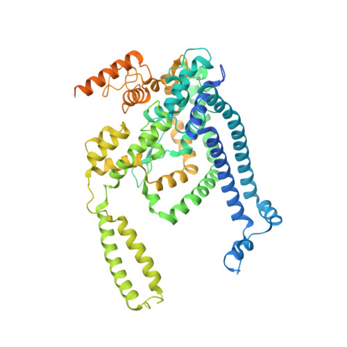
Structure of Human ATG9A, the Only Transmembrane Protein of the Core Autophagy Machinery.

Guardia, C.M.Tan, X.F.Lian, T.Rana, M.S.Zhou, W.Christenson, E.T.Lowry, A.J.Faraldo-Gomez, J.D.Bonifacino, J.S.Jiang, J.Banerjee, A.

(2020) Cell Rep 31: 107837-107837

  • DOI: https://doi.org/10.1016/j.celrep.2020.107837
  • Primary Citation of Related Structures:  
    6WQZ, 6WR4

  • PubMed Abstract: 
  • Autophagy is a catabolic process involving capture of cytoplasmic materials into double-membraned autophagosomes that subsequently fuse with lysosomes for degradation of the materials by lysosomal hydrolases. One of the least understood components of the autophagy machinery is the transmembrane protein ATG9 ...

    Autophagy is a catabolic process involving capture of cytoplasmic materials into double-membraned autophagosomes that subsequently fuse with lysosomes for degradation of the materials by lysosomal hydrolases. One of the least understood components of the autophagy machinery is the transmembrane protein ATG9. Here, we report a cryoelectron microscopy structure of the human ATG9A isoform at 2.9-Å resolution. The structure reveals a fold with a homotrimeric domain-swapped architecture, multiple membrane spans, and a network of branched cavities, consistent with ATG9A being a membrane transporter. Mutational analyses support a role for the cavities in the function of ATG9A. In addition, structure-guided molecular simulations predict that ATG9A causes membrane bending, explaining the localization of this protein to small vesicles and highly curved edges of growing autophagosomes.


    Organizational Affiliation

    Unit on Structural and Chemical Biology of Membrane Proteins, Neurosciences and Cellular and Structural Biology Division, Eunice Kennedy Shriver National Institute of Child Health and Human Development, National Institutes of Health, Bethesda, MD 20892, USA. Electronic address: anirban.banerjee@nih.gov.


Macromolecules
Find similar proteins by:  (by identity cutoff)  |  3D Structure
Entity ID: 1
MoleculeChainsSequence LengthOrganismDetailsImage
Autophagy-related protein 9A724Homo sapiensMutation(s): 0 
Gene Names: ATG9AAPG9L1
Membrane Entity: Yes 
UniProt & NIH Common Fund Data Resources
Find proteins for Q7Z3C6 (Homo sapiens)
Explore Q7Z3C6 
Go to UniProtKB:  Q7Z3C6
PHAROS:  Q7Z3C6
Entity Groups  
Sequence Clusters30% Identity50% Identity70% Identity90% Identity95% Identity100% Identity
UniProt GroupQ7Z3C6
Protein Feature View
Expand
  • Reference Sequence
Small Molecules
Ligands 1 Unique
IDChainsName / Formula / InChI Key2D Diagram3D Interactions
LMN
Query on LMN

Download Ideal Coordinates CCD File 
G [auth C],
H [auth C],
I [auth B],
J [auth B],
K [auth A],
G [auth C],
H [auth C],
I [auth B],
J [auth B],
K [auth A],
L [auth A]
Lauryl Maltose Neopentyl Glycol
C47 H88 O22
MADJBYLAYPCCOO-XYPZXBMFSA-N
 Ligand Interaction
Experimental Data & Validation

Experimental Data

  • Method: ELECTRON MICROSCOPY
  • Resolution: 2.80 Å
  • Aggregation State: PARTICLE 
  • Reconstruction Method: SINGLE PARTICLE 

Structure Validation

View Full Validation Report


Entry History 

Revision History  (Full details and data files)

  • Version 1.0: 2020-07-08
    Type: Initial release
  • Version 1.1: 2020-07-15
    Changes: Database references