6WPH

Structure of HIV-1 Reverse Transcriptase (RT) in complex with dsDNA and (-)-FTC


Experimental Data Snapshot

  • Method: X-RAY DIFFRACTION
  • Resolution: 2.72 Å
  • R-Value Free: 0.257 
  • R-Value Work: 0.197 
  • R-Value Observed: 0.200 

wwPDB Validation   3D Report Full Report


Ligand Structure Quality Assessment 


This is version 1.1 of the entry. See complete history


Literature

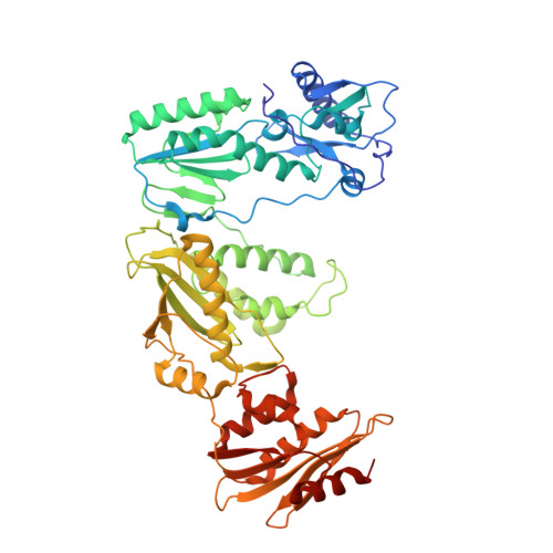
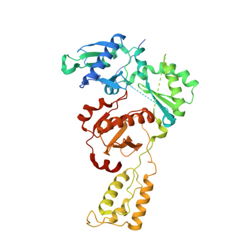
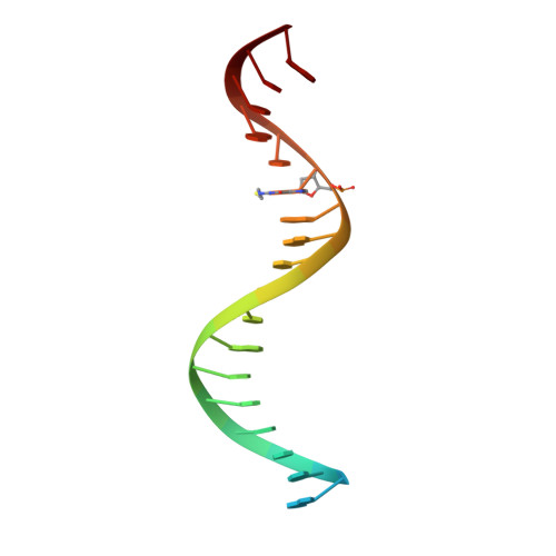
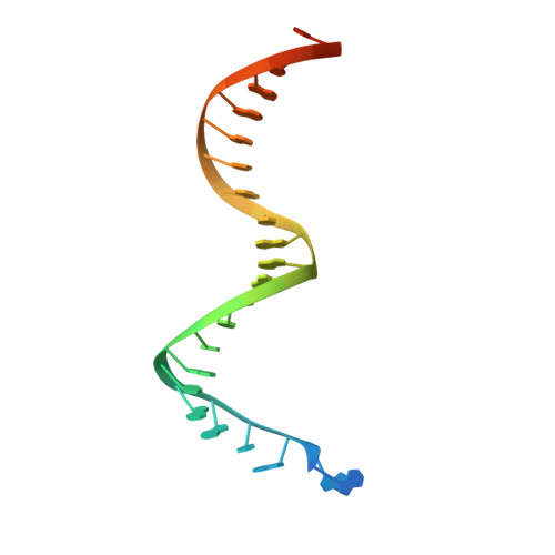
Post-Catalytic Complexes with Emtricitabine or Stavudine and HIV-1 Reverse Transcriptase Reveal New Mechanistic Insights for Nucleotide Incorporation and Drug Resistance.

Bertoletti, N.Chan, A.H.Schinazi, R.F.Anderson, K.S.

(2020) Molecules 25

  • DOI: https://doi.org/10.3390/molecules25204868
  • Primary Citation of Related Structures:  
    6WPF, 6WPH, 6WPJ

  • PubMed Abstract: 

    Human immunodeficiency virus 1 (HIV-1) infection is a global health issue since neither a cure nor a vaccine is available. However, the highly active antiretroviral therapy (HAART) has improved the life expectancy for patients with acquired immunodeficiency syndrome (AIDS). Nucleoside reverse transcriptase inhibitors (NRTIs) are in almost all HAART and target reverse transcriptase (RT), an essential enzyme for the virus. Even though NRTIs are highly effective, they have limitations caused by RT resistance. The main mechanisms of RT resistance to NRTIs are discrimination and excision. Understanding the molecular mechanisms for discrimination and excision are essential to develop more potent and selective NRTIs. Using protein X-ray crystallography, we determined the first crystal structure of RT in its post-catalytic state in complex with emtricitabine, (-)FTC or stavudine (d4T). Our structural studies provide the framework for understanding how RT discriminates between NRTIs and natural nucleotides, and for understanding the requirement of (-)FTC to undergo a conformation change for successful incorporation by RT. The crystal structure of RT in post-catalytic complex with d4T provides a "snapshot" for considering the possible mechanism of how RT develops resistance for d4T via excision. The findings reported herein will contribute to the development of next generation NRTIs.


  • Organizational Affiliation

    Department of Pharmacology, Yale University School of Medicine, New Haven, CT 06520-8066, USA.


Macromolecules

Find similar proteins by:  (by identity cutoff)  |  3D Structure
Entity ID: 1
MoleculeChains Sequence LengthOrganismDetailsImage
Reverse transcriptase/ribonuclease H560HIV-1 M:B_HXB2RMutation(s): 2 
Gene Names: gag-pol
EC: 2.7.7.49 (PDB Primary Data), 2.7.7.7 (PDB Primary Data), 3.1.26.13 (PDB Primary Data)
UniProt
Find proteins for P04585 (Human immunodeficiency virus type 1 group M subtype B (isolate HXB2))
Explore P04585 
Go to UniProtKB:  P04585
Entity Groups  
Sequence Clusters30% Identity50% Identity70% Identity90% Identity95% Identity100% Identity
UniProt GroupP04585
Sequence Annotations
Expand
  • Reference Sequence
Find similar proteins by:  (by identity cutoff)  |  3D Structure
Entity ID: 2
MoleculeChains Sequence LengthOrganismDetailsImage
p51 RT452HIV-1 M:B_HXB2RMutation(s): 1 
Gene Names: gag-pol
EC: 2.7.7 (PDB Primary Data), 3.1 (PDB Primary Data)
UniProt
Find proteins for P04585 (Human immunodeficiency virus type 1 group M subtype B (isolate HXB2))
Explore P04585 
Go to UniProtKB:  P04585
Entity Groups  
Sequence Clusters30% Identity50% Identity70% Identity90% Identity95% Identity100% Identity
UniProt GroupP04585
Sequence Annotations
Expand
  • Reference Sequence

Find similar nucleic acids by:  Sequence   |   3D Structure  

Entity ID: 3
MoleculeChains LengthOrganismImage
DNA Primer 21-merC [auth P]21synthetic construct
Sequence Annotations
Expand
  • Reference Sequence
Find similar nucleic acids by:  (by identity cutoff)  |  3D Structure
Entity ID: 4
MoleculeChains LengthOrganismImage
DNA template 27-merD [auth T]27synthetic construct
Sequence Annotations
Expand
  • Reference Sequence
Small Molecules
Ligands 4 Unique
IDChains Name / Formula / InChI Key2D Diagram3D Interactions
DG
Query on DG

Download Ideal Coordinates CCD File 
I [auth T]2'-DEOXYGUANOSINE-5'-MONOPHOSPHATE
C10 H14 N5 O7 P
LTFMZDNNPPEQNG-KVQBGUIXSA-N
43X (Subject of Investigation/LOI)
Query on 43X

Download Ideal Coordinates CCD File 
G [auth P][(2R,5S)-5-(4-amino-5-fluoro-2-oxopyrimidin-1(2H)-yl)-1,3-oxathiolan-2-yl]methyl dihydrogen phosphate
C8 H11 F N3 O6 P S
WQOMZMVXTRGMHZ-NTSWFWBYSA-N
SO4
Query on SO4

Download Ideal Coordinates CCD File 
H [auth T],
J [auth T]
SULFATE ION
O4 S
QAOWNCQODCNURD-UHFFFAOYSA-L
MG
Query on MG

Download Ideal Coordinates CCD File 
E [auth A],
F [auth B]
MAGNESIUM ION
Mg
JLVVSXFLKOJNIY-UHFFFAOYSA-N
Experimental Data & Validation

Experimental Data

  • Method: X-RAY DIFFRACTION
  • Resolution: 2.72 Å
  • R-Value Free: 0.257 
  • R-Value Work: 0.197 
  • R-Value Observed: 0.200 
  • Space Group: C 2 2 21
Unit Cell:
Length ( Å )Angle ( ˚ )
a = 165.98α = 90
b = 168.054β = 90
c = 101.916γ = 90
Software Package:
Software NamePurpose
XDSdata scaling
PHENIXrefinement
XDSdata reduction
PHASERphasing

Structure Validation

View Full Validation Report


Ligand Structure Quality Assessment 


Entry History & Funding Information

Deposition Data


Funding OrganizationLocationGrant Number
National Institutes of Health/National Institute of General Medical Sciences (NIH/NIGMS)United StatesGM049551

Revision History  (Full details and data files)

  • Version 1.0: 2020-11-04
    Type: Initial release
  • Version 1.1: 2023-10-18
    Changes: Data collection, Database references, Refinement description