Structural basis for substrate recognition and chemical inhibition of oncogenic MAGE ubiquitin ligases.
Yang, S.W., Huang, X., Lin, W., Min, J., Miller, D.J., Mayasundari, A., Rodrigues, P., Griffith, E.C., Gee, C.T., Li, L., Li, W., Lee, R.E., Rankovic, Z., Chen, T., Potts, P.R.(2020) Nat Commun 11: 4931-4931
- PubMed: 33004795 Search on PubMedSearch on PubMed Central
- DOI: https://doi.org/10.1038/s41467-020-18708-x
- Primary Citation Related Structures:
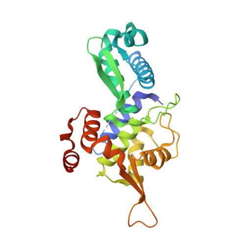
6WJH - PubMed Abstract:
Testis-restricted melanoma antigen (MAGE) proteins are frequently hijacked in cancer and play a critical role in tumorigenesis. MAGEs assemble with E3 ubiquitin ligases and function as substrate adaptors that direct the ubiquitination of novel targets, including key tumor suppressors. However, how MAGEs recognize their targets is unknown and has impeded the development of MAGE-directed therapeutics. Here, we report the structural basis for substrate recognition by MAGE ubiquitin ligases. Biochemical analysis of the degron motif recognized by MAGE-A11 and the crystal structure of MAGE-A11 bound to the PCF11 substrate uncovered a conserved substrate binding cleft (SBC) in MAGEs. Mutation of the SBC disrupted substrate recognition by MAGEs and blocked MAGE-A11 oncogenic activity. A chemical screen for inhibitors of MAGE-A11:substrate interaction identified 4-Aminoquinolines as potent inhibitors of MAGE-A11 that show selective cytotoxicity. These findings provide important insights into the large family of MAGE ubiquitin ligases and identify approaches for developing cancer-specific therapeutics.
- Department of Cell and Molecular Biology, St. Jude Children's Research Hospital, 262 Danny Thomas Pl, Memphis, TN, 38105, USA.
Organizational Affiliation: