6W51 | pdb_00006w51

Structure of the antibody fragment H2 in complex with HLA-A*02:01/p53R175H


Experimental Data Snapshot

  • Method: X-RAY DIFFRACTION
  • Resolution: 3.53 Å
  • R-Value Free: 
    0.283 (Depositor), 0.277 (DCC) 
  • R-Value Work: 
    0.200 (Depositor), 0.201 (DCC) 
  • R-Value Observed: 
    0.204 (Depositor) 

Starting Models: experimental
View more details

wwPDB Validation 3D Report Full Report

Validation slider image for 6W51

This is version 1.2 of the entry. See complete history

Literature

Targeting a neoantigen derived from a common TP53 mutation.

Hsiue, E.H.Wright, K.M.Douglass, J.Hwang, M.S.Mog, B.J.Pearlman, A.H.Paul, S.DiNapoli, S.R.Konig, M.F.Wang, Q.Schaefer, A.Miller, M.S.Skora, A.D.Azurmendi, P.A.Murphy, M.B.Liu, Q.Watson, E.Li, Y.Pardoll, D.M.Bettegowda, C.Papadopoulos, N.Kinzler, K.W.Vogelstein, B.Gabelli, S.B.Zhou, S.

(2021) Science 371

  • DOI: https://doi.org/10.1126/science.abc8697
  • Primary Citation Related Structures: 
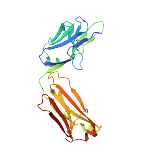
    6W51

  • PubMed Abstract: 

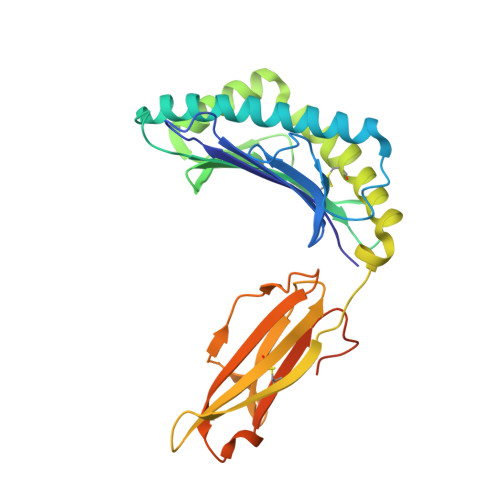
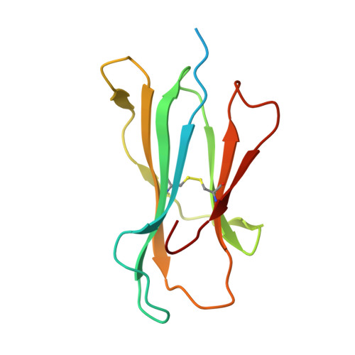
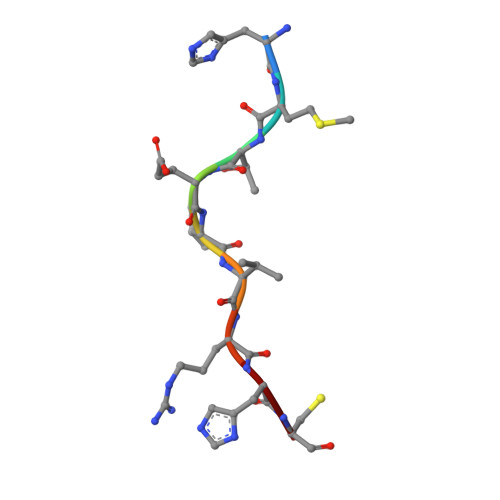
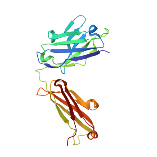
    TP53 (tumor protein p53) is the most commonly mutated cancer driver gene, but drugs that target mutant tumor suppressor genes, such as TP53 , are not yet available. Here, we describe the identification of an antibody highly specific to the most common TP53 mutation (R175H, in which arginine at position 175 is replaced with histidine) in complex with a common human leukocyte antigen-A (HLA-A) allele on the cell surface. We describe the structural basis of this specificity and its conversion into an immunotherapeutic agent: a bispecific single-chain diabody. Despite the extremely low p53 peptide-HLA complex density on the cancer cell surface, the bispecific antibody effectively activated T cells to lyse cancer cells that presented the neoantigen in vitro and in mice. This approach could in theory be used to target cancers containing mutations that are difficult to target in conventional ways.


  • Organizational Affiliation
    • Ludwig Center, Sidney Kimmel Comprehensive Cancer Center, Johns Hopkins University School of Medicine, Baltimore, MD 21287, USA.

Macromolecule Content 

  • Total Structure Weight: 386.13 kDa 
  • Atom Count: 25,900 
  • Modeled Residue Count: 3,264 
  • Deposited Residue Count: 3,448 
  • Unique protein chains: 5

Macromolecules

Find similar proteins by:|  3D Structure
Entity ID: 1
MoleculeChains  Sequence LengthOrganismDetailsImage
MHC class I antigen
A, D, G, J
296Homo sapiensMutation(s): 0 
Gene Names: HLA-A
UniProt
Find proteins for A0A140T913 (Homo sapiens)
Explore A0A140T913 
Go to UniProtKB:  A0A140T913
Entity Groups
Sequence Clusters30% Identity50% Identity70% Identity90% Identity95% Identity100% Identity
UniProt GroupA0A140T913
Sequence Annotations
Expand
Reference Sequence
Find similar proteins by:|  3D Structure
Entity ID: 2
MoleculeChains  Sequence LengthOrganismDetailsImage
Beta-2-microglobulin
B, E, H, K
119Homo sapiensMutation(s): 0 
Gene Names: B2MCDABP0092HDCMA22P
UniProt & NIH Common Fund Data Resources
Find proteins for P61769 (Homo sapiens)
Explore P61769 
Go to UniProtKB:  P61769
PHAROS:  P61769
GTEx:  ENSG00000166710 
Entity Groups
Sequence Clusters30% Identity50% Identity70% Identity90% Identity95% Identity100% Identity
UniProt GroupP61769
Sequence Annotations
Expand
Reference Sequence
Find similar proteins by:  Sequence   |   3D Structure  
Entity ID: 3
MoleculeChains  Sequence LengthOrganismDetailsImage
Cellular tumor antigen p53 peptide
C, F, I, L
9Homo sapiensMutation(s): 1 
UniProt & NIH Common Fund Data Resources
Find proteins for P04637 (Homo sapiens)
Explore P04637 
Go to UniProtKB:  P04637
PHAROS:  P04637
GTEx:  ENSG00000141510 
Entity Groups
UniProt GroupP04637
Sequence Annotations
Expand
Reference Sequence
Find similar proteins by:|  3D Structure
Entity ID: 4
MoleculeChains  Sequence LengthOrganismDetailsImage
Immunoglobulin heavy chain H2
M, O, Q, S
223Homo sapiensMutation(s): 0 
Find similar proteins by:|  3D Structure
Entity ID: 5
MoleculeChains  Sequence LengthOrganismDetailsImage
Immunoglobulin light chain H2
N, P, R, T
215Homo sapiensMutation(s): 0 

Small Molecules

Ligands 1 Unique
IDChains Name / Formula / InChI Key2D Diagram3D Interactions
PO4

Query on PO4


Download:Ideal Coordinates CCD File
U [auth S]PHOSPHATE ION
O4 P
NBIIXXVUZAFLBC-UHFFFAOYSA-K

Experimental Data & Validation

Experimental Data

  • Method: X-RAY DIFFRACTION
  • Resolution: 3.53 Å
  • R-Value Free:  0.283 (Depositor), 0.277 (DCC) 
  • R-Value Work:  0.200 (Depositor), 0.201 (DCC) 
  • R-Value Observed: 0.204 (Depositor) 
Space Group: P 1 21 1
Unit Cell:
Length ( Å )Angle ( ˚ )
a = 113.21α = 90
b = 123.69β = 100.36
c = 136.909γ = 90
Software Package:
Software NamePurpose
REFMACrefinement
XDSdata reduction
Aimlessdata scaling
PDB_EXTRACTdata extraction
PHASERphasing

Structure Validation

View Full Validation Report


Entry History 

Deposition Data

Revision History  (Full details and data files)

  • Version 1.0: 2021-03-17
    Type: Initial release
  • Version 1.1: 2023-10-18
    Changes: Data collection, Database references, Refinement description
  • Version 1.2: 2024-11-20
    Changes: Structure summary