







 Download Mendeley
Co-crystal structure of Pd_dinase with probe glycine-propargylglycine-AOMK
Xu, J.H., Solania, A., Wolan, D.W.To be published.
Experimental Data Snapshot
Starting Model: experimental
View more details
Entity ID: 1 | |||||
|---|---|---|---|---|---|
| Molecule | Chains | Sequence Length | Organism | Details | Image |
| Aminopeptidase | 401 | Parabacteroides distasonis | Mutation(s): 0 Gene Names: DW188_02780, DW692_01025, DW700_01025, DW808_17970, DW814_08450, DW867_06285, DW984_01045, DW998_13650, DWW19_04025, DWY54_02375... | 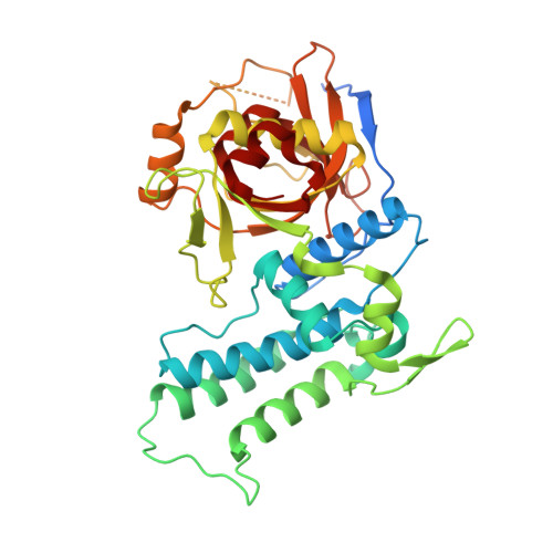 | |
UniProt | |||||
Entity Groups | |||||
| Sequence Clusters | 30% Identity50% Identity70% Identity90% Identity95% Identity100% Identity | ||||
| UniProt Group | A6LE66 | ||||
Sequence AnnotationsExpand | |||||
Reference Sequence | |||||
| Ligands 2 Unique | |||||
|---|---|---|---|---|---|
| ID | Chains | Name / Formula / InChI Key | 2D Diagram | 3D Interactions | |
| SWJ (Subject of Investigation/LOI) Download:Ideal Coordinates CCD File | G [auth A] J [auth B] M [auth C] P [auth D] S [auth E] | 2-azanyl-~{N}-[(3~{S})-2-oxidanylidenehex-5-yn-3-yl]ethanamide C8 H12 N2 O2 YHGRTOPUPRQLPX-ZETCQYMHSA-N | 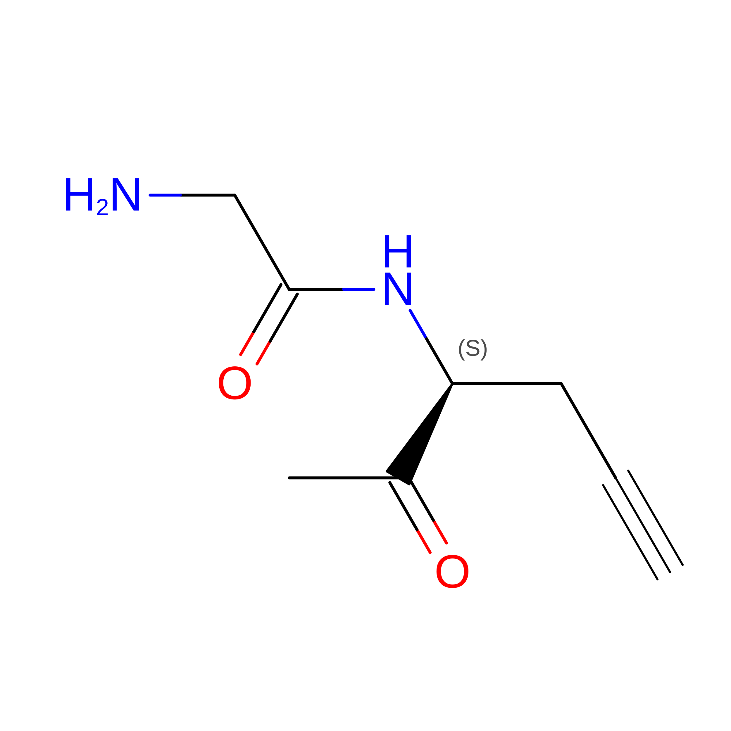 | ||
| K Download:Ideal Coordinates CCD File | H [auth A] I [auth A] K [auth B] L [auth B] N [auth C] | POTASSIUM ION K NPYPAHLBTDXSSS-UHFFFAOYSA-N |  | ||
| Length ( Å ) | Angle ( ˚ ) |
|---|---|
| a = 105.113 | α = 90 |
| b = 138.613 | β = 90 |
| c = 220.13 | γ = 90 |
| Software Name | Purpose |
|---|---|
| PHENIX | refinement |
| HKL-2000 | data scaling |
| PDB_EXTRACT | data extraction |
| HKL-2000 | data reduction |
| PHASER | phasing |








