6VQJ

Mammalian V-ATPase from rat brain collar and peripheral stalks rotational state 2 (from focused refinement)


Experimental Data Snapshot

  • Method: ELECTRON MICROSCOPY
  • Resolution: 5.70 Å
  • Aggregation State: PARTICLE 
  • Reconstruction Method: SINGLE PARTICLE 

wwPDB Validation   3D Report Full Report


This is version 1.2 of the entry. See complete history


Literature

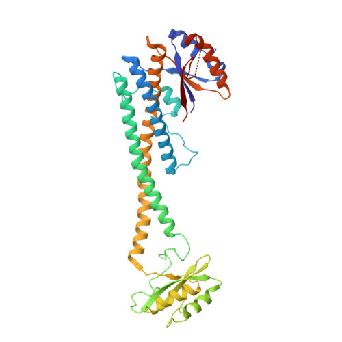
Structure of V-ATPase from the mammalian brain.

Abbas, Y.M.Wu, D.Bueler, S.A.Robinson, C.V.Rubinstein, J.L.

(2020) Science 367: 1240-1246

  • DOI: https://doi.org/10.1126/science.aaz2924
  • Primary Citation of Related Structures:  
    6VQ6, 6VQ7, 6VQ8, 6VQ9, 6VQA, 6VQB, 6VQC, 6VQG, 6VQH, 6VQI, 6VQJ, 6VQK

  • PubMed Abstract: 

    In neurons, the loading of neurotransmitters into synaptic vesicles uses energy from proton-pumping vesicular- or vacuolar-type adenosine triphosphatases (V-ATPases). These membrane protein complexes possess numerous subunit isoforms, which complicates their analysis. We isolated homogeneous rat brain V-ATPase through its interaction with SidK, a Legionella pneumophila effector protein. Cryo-electron microscopy allowed the construction of an atomic model, defining the enzyme's ATP:proton ratio as 3:10 and revealing a homolog of yeast subunit f in the membrane region, which we tentatively identify as RNAseK. The c ring encloses the transmembrane anchors for cleaved ATP6AP1/Ac45 and ATP6AP2/PRR, the latter of which is the (pro)renin receptor that, in other contexts, is involved in both Wnt signaling and the renin-angiotensin system that regulates blood pressure. This structure shows how ATP6AP1/Ac45 and ATP6AP2/PRR enable assembly of the enzyme's catalytic and membrane regions.


  • Organizational Affiliation

    Molecular Medicine Program, The Hospital for Sick Children Research Institute, Toronto, ON M5G 0A4, Canada.


Macromolecules
Find similar proteins by:  (by identity cutoff)  |  3D Structure
Entity ID: 1
MoleculeChains Sequence LengthOrganismDetailsImage
V-type proton ATPase subunit C 1A [auth G]382Rattus norvegicusMutation(s): 0 
UniProt
Find proteins for Q5FVI6 (Rattus norvegicus)
Explore Q5FVI6 
Go to UniProtKB:  Q5FVI6
Entity Groups  
Sequence Clusters30% Identity50% Identity70% Identity90% Identity95% Identity100% Identity
UniProt GroupQ5FVI6
Sequence Annotations
Expand
  • Reference Sequence
Find similar proteins by:  (by identity cutoff)  |  3D Structure
Entity ID: 2
MoleculeChains Sequence LengthOrganismDetailsImage
V-type proton ATPase subunit E 1B [auth I],
C [auth J],
D [auth K]
226Rattus norvegicusMutation(s): 0 
UniProt
Find proteins for Q6PCU2 (Rattus norvegicus)
Explore Q6PCU2 
Go to UniProtKB:  Q6PCU2
Entity Groups  
Sequence Clusters30% Identity50% Identity70% Identity90% Identity95% Identity100% Identity
UniProt GroupQ6PCU2
Sequence Annotations
Expand
  • Reference Sequence
Find similar proteins by:  (by identity cutoff)  |  3D Structure
Entity ID: 3
MoleculeChains Sequence LengthOrganismDetailsImage
V-type proton ATPase subunit GE [auth M],
F [auth N],
G [auth O]
118Rattus norvegicusMutation(s): 0 
UniProt
Find proteins for Q8R2H0 (Rattus norvegicus)
Explore Q8R2H0 
Go to UniProtKB:  Q8R2H0
Entity Groups  
Sequence Clusters30% Identity50% Identity70% Identity90% Identity95% Identity100% Identity
UniProt GroupQ8R2H0
Sequence Annotations
Expand
  • Reference Sequence
Find similar proteins by:  (by identity cutoff)  |  3D Structure
Entity ID: 4
MoleculeChains Sequence LengthOrganismDetailsImage
V-type proton ATPase 116 kDa subunit a isoform 1H [auth a]838Rattus norvegicusMutation(s): 0 
UniProt
Find proteins for P25286 (Rattus norvegicus)
Explore P25286 
Go to UniProtKB:  P25286
Entity Groups  
Sequence Clusters30% Identity50% Identity70% Identity90% Identity95% Identity100% Identity
UniProt GroupP25286
Sequence Annotations
Expand
  • Reference Sequence
Experimental Data & Validation

Experimental Data

  • Method: ELECTRON MICROSCOPY
  • Resolution: 5.70 Å
  • Aggregation State: PARTICLE 
  • Reconstruction Method: SINGLE PARTICLE 
EM Software:
TaskSoftware PackageVersion
RECONSTRUCTIONcryoSPARC

Structure Validation

View Full Validation Report


Entry History & Funding Information

Deposition Data


Funding OrganizationLocationGrant Number
Canadian Institutes of Health Research (CIHR)Canada--

Revision History  (Full details and data files)

  • Version 1.0: 2020-03-18
    Type: Initial release
  • Version 1.1: 2020-09-30
    Changes: Database references
  • Version 1.2: 2024-03-06
    Changes: Data collection, Database references