6VPQ

Crystal structure of the C-terminal domain of DENR


Experimental Data Snapshot

  • Method: X-RAY DIFFRACTION
  • Resolution: 1.74 Å
  • R-Value Free: 0.247 
  • R-Value Work: 0.178 
  • R-Value Observed: 0.181 

wwPDB Validation   3D Report Full Report


This is version 1.3 of the entry. See complete history


Literature

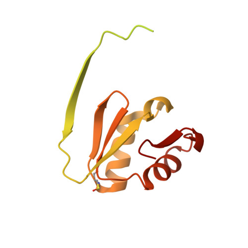
Crystal structure of the C-terminal domain of DENR.

Lomakin, I.B.De, S.Wang, J.Borkar, A.N.Steitz, T.A.

(2020) Comput Struct Biotechnol J 18: 696-704

  • DOI: https://doi.org/10.1016/j.csbj.2020.03.009
  • Primary Citation of Related Structures:  
    6VPQ, 6VPR

  • PubMed Abstract: 

    The density regulated protein (DENR) forms a stable heterodimer with malignant T-cell-amplified sequence 1 (MCT-1). DENR-MCT-1 heterodimer then participates in regulation of non-canonical translation initiation and ribosomal recycling. The N-terminal domain of DENR interacts with MCT-1 and carries a classical tetrahedral zinc ion-binding site, which is crucial for the dimerization. DENR-MCT-1 binds the small (40S) ribosomal subunit through interactions between MCT-1 and helix h24 of the 18S rRNA, and through interactions between the C-terminal domain of DENR and helix h44 of the 18S rRNA. This later interaction occurs in the vicinity of the P site that is also the binding site for canonical translation initiation factor eIF1, which plays the key role in initiation codon selection and scanning. Sequence homology modeling and a low-resolution crystal structure of the DENR-MCT-1 complex with the human 40S subunit suggests that the C-terminal domain of DENR and eIF1 adopt a similar fold. Here we present the crystal structure of the C-terminal domain of DENR determined at 1.74 Å resolution, which confirms its resemblance to eIF1 and advances our understanding of the mechanism by which DENR-MCT-1 regulates non-canonical translation initiation and ribosomal recycling.


  • Organizational Affiliation

    Department of Molecular Biophysics and Biochemistry, Yale University, New Haven, CT 06520-8114, USA.


Macromolecules
Find similar proteins by:  (by identity cutoff)  |  3D Structure
Entity ID: 1
MoleculeChains Sequence LengthOrganismDetailsImage
Density-regulated protein
A, B, C, D, E
A, B, C, D, E, F
198Homo sapiensMutation(s): 0 
Gene Names: DENRDRP1H14
UniProt & NIH Common Fund Data Resources
Find proteins for O43583 (Homo sapiens)
Explore O43583 
Go to UniProtKB:  O43583
PHAROS:  O43583
GTEx:  ENSG00000139726 
Entity Groups  
Sequence Clusters30% Identity50% Identity70% Identity90% Identity95% Identity100% Identity
UniProt GroupO43583
Sequence Annotations
Expand
  • Reference Sequence
Small Molecules
Ligands 4 Unique
IDChains Name / Formula / InChI Key2D Diagram3D Interactions
PGE
Query on PGE

Download Ideal Coordinates CCD File 
CA [auth D]
IA [auth E]
J [auth A]
K [auth A]
QA [auth F]
CA [auth D],
IA [auth E],
J [auth A],
K [auth A],
QA [auth F],
RA [auth F],
S [auth B],
T [auth B],
W [auth C],
X [auth C],
Y [auth C]
TRIETHYLENE GLYCOL
C6 H14 O4
ZIBGPFATKBEMQZ-UHFFFAOYSA-N
ACT
Query on ACT

Download Ideal Coordinates CCD File 
M [auth A],
SA [auth F],
TA [auth F]
ACETATE ION
C2 H3 O2
QTBSBXVTEAMEQO-UHFFFAOYSA-M
CA
Query on CA

Download Ideal Coordinates CCD File 
AA [auth C],
EA [auth D],
U [auth B]
CALCIUM ION
Ca
BHPQYMZQTOCNFJ-UHFFFAOYSA-N
MG
Query on MG

Download Ideal Coordinates CCD File 
BA [auth D]
DA [auth D]
FA [auth E]
G [auth A]
GA [auth E]
BA [auth D],
DA [auth D],
FA [auth E],
G [auth A],
GA [auth E],
H [auth A],
HA [auth E],
I [auth A],
JA [auth F],
KA [auth F],
L [auth A],
LA [auth F],
MA [auth F],
N [auth B],
NA [auth F],
O [auth B],
OA [auth F],
P [auth B],
PA [auth F],
Q [auth B],
R [auth B],
V [auth C],
Z [auth C]
MAGNESIUM ION
Mg
JLVVSXFLKOJNIY-UHFFFAOYSA-N
Modified Residues  1 Unique
IDChains TypeFormula2D DiagramParent
CSO
Query on CSO
A, B, C, D, E
A, B, C, D, E, F
L-PEPTIDE LINKINGC3 H7 N O3 SCYS
Experimental Data & Validation

Experimental Data

  • Method: X-RAY DIFFRACTION
  • Resolution: 1.74 Å
  • R-Value Free: 0.247 
  • R-Value Work: 0.178 
  • R-Value Observed: 0.181 
  • Space Group: P 1 21 1
Unit Cell:
Length ( Å )Angle ( ˚ )
a = 64.67α = 90
b = 47.26β = 106.29
c = 95.5γ = 90
Software Package:
Software NamePurpose
XDSdata reduction
XSCALEdata scaling
PHENIXrefinement
PDB_EXTRACTdata extraction
PHASERphasing

Structure Validation

View Full Validation Report


Entry History & Funding Information

Deposition Data


Funding OrganizationLocationGrant Number
Howard Hughes Medical Institute (HHMI)United States--
National Institutes of Health/National Institute of General Medical Sciences (NIH/NIGMS)United StatesGM022778

Revision History  (Full details and data files)

  • Version 1.0: 2020-04-08
    Type: Initial release
  • Version 1.1: 2020-04-22
    Changes: Database references
  • Version 1.2: 2023-10-11
    Changes: Advisory, Data collection, Database references, Derived calculations, Refinement description
  • Version 1.3: 2023-11-15
    Changes: Data collection