6V8J | pdb_00006v8j

Crystal structure of Ara h 8.0201


Experimental Data Snapshot

  • Method: X-RAY DIFFRACTION
  • Resolution: 1.95 Å
  • R-Value Free: 
    0.219 (Depositor), 0.254 (DCC) 
  • R-Value Work: 
    0.173 (Depositor), 0.207 (DCC) 
  • R-Value Observed: 
    0.175 (Depositor) 

Starting Model: experimental
View more details

wwPDB Validation   3D Report Full Report


This is version 1.2 of the entry. See complete history


Literature

Structural analysis of 8-anilino-1-naphthalene sulfonate (ANS) binding to the PR-10 allergen Ara h 8.

O'Malley, A.Offermann, L.R.Khatri, K.Linn, C.Pote, S.McBride, J.K.Perdue, M.L.Hurlburt, B.K.Maleki, S.J.Mias, G.I.Chruszcz, M.

(2025) Biochem Biophys Res Commun 793: 153013-153013

  • DOI: https://doi.org/10.1016/j.bbrc.2025.153013
  • Primary Citation of Related Structures:  
    6AWR, 6V8H, 6V8J, 6V8M, 6V8S

  • PubMed Abstract: 

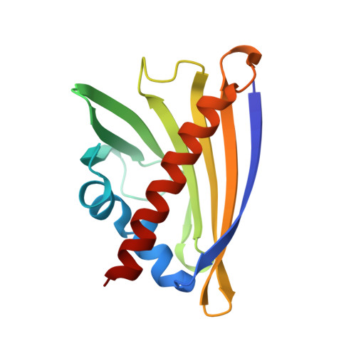
    We previously determined crystal structures of peanut allergen Ara h 8.0101 in the apo form as well as in complex with model ligands. These structures illustrated the varied ligand binding capabilities of PR-10s and Ara h 8's structural similarity to the major birch allergen Bet v 1. Here, we expanded on those structural studies with structures of Ara h 8.0101 and Ara h 8.0201 in complex with 8-anilino-1-naphthalene sulfonate (ANS), as well as the apo form of Ara h 8.0201. Structural studies revealed that both proteins may bind more than one ANS molecule. We also examined the impact of ANS on the ligand binding cavities of Ara h 8.0101 and Ara h 8.0201 with fluorescence assays and compared the results to prototypic PR-10 Bet v 1.0101. Moreover, as ANS is often used in fluorescence-based ligand binding assays, we analyzed structures from the PDB and provided a summary on experimentally determined ANS binding sites. These analyses show that ANS is useful for investigation of ligand binding sites, but it may also participate in non-specific reactions on nonpolar surfaces of proteins.


  • Organizational Affiliation
    • Department of Biochemistry and Molecular Biology, Michigan State University, East Lansing, MI, 48824, USA.

Macromolecules
Find similar proteins by:  (by identity cutoff)  |  3D Structure
Entity ID: 1
MoleculeChains Sequence LengthOrganismDetailsImage
Ara h 8 allergen isoform
A, B, C, D
153Arachis hypogaeaMutation(s): 0 
Gene Names: Ahy_Scaffold6g108211
UniProt
Find proteins for B0YIU5 (Arachis hypogaea)
Explore B0YIU5 
Go to UniProtKB:  B0YIU5
Entity Groups  
Sequence Clusters30% Identity50% Identity70% Identity90% Identity95% Identity100% Identity
UniProt GroupB0YIU5
Sequence Annotations
Expand
  • Reference Sequence
Small Molecules
Ligands 2 Unique
IDChains Name / Formula / InChI Key2D Diagram3D Interactions
SO4
Query on SO4

Download Ideal Coordinates CCD File 
F [auth A]
G [auth A]
H [auth A]
J [auth B]
L [auth C]
F [auth A],
G [auth A],
H [auth A],
J [auth B],
L [auth C],
M [auth C],
N [auth C],
P [auth D]
SULFATE ION
O4 S
QAOWNCQODCNURD-UHFFFAOYSA-L
NA
Query on NA

Download Ideal Coordinates CCD File 
E [auth A],
I [auth B],
K [auth C],
O [auth D]
SODIUM ION
Na
FKNQFGJONOIPTF-UHFFFAOYSA-N
Experimental Data & Validation

Experimental Data

  • Method: X-RAY DIFFRACTION
  • Resolution: 1.95 Å
  • R-Value Free:  0.219 (Depositor), 0.254 (DCC) 
  • R-Value Work:  0.173 (Depositor), 0.207 (DCC) 
  • R-Value Observed: 0.175 (Depositor) 
Space Group: P 1
Unit Cell:
Length ( Å )Angle ( ˚ )
a = 38.406α = 89.96
b = 59.209β = 89.99
c = 67.078γ = 71.02
Software Package:
Software NamePurpose
REFMACrefinement
PDB_EXTRACTdata extraction
HKL-2000data reduction
HKL-2000data scaling
MOLREPphasing
Cootmodel building
BUCCANEERmodel building

Structure Validation

View Full Validation Report


Entry History 

Deposition Data

Revision History  (Full details and data files)

  • Version 1.0: 2020-12-16
    Type: Initial release
  • Version 1.1: 2023-10-11
    Changes: Data collection, Database references, Refinement description
  • Version 1.2: 2025-12-03
    Changes: Database references, Structure summary