6V5V | pdb_00006v5v

Structure of gamma-tubulin in the native human gamma-tubulin ring complex


Experimental Data Snapshot

  • Method: ELECTRON MICROSCOPY
  • Resolution: 3.80 Å
  • Aggregation State: PARTICLE 
  • Reconstruction Method: SINGLE PARTICLE 

wwPDB Validation 3D Report Full Report

Validation slider image for 6V5V

This is version 1.2 of the entry. See complete history

Literature

Asymmetric Molecular Architecture of the Human gamma-Tubulin Ring Complex.

Wieczorek, M.Urnavicius, L.Ti, S.C.Molloy, K.R.Chait, B.T.Kapoor, T.M.

(2020) Cell 180: 165-175.e16

  • DOI: https://doi.org/10.1016/j.cell.2019.12.007
  • Primary Citation Related Structures: 
    6V5V, 6V69, 6V6B, 6V6C, 6V6S

  • PubMed Abstract: 

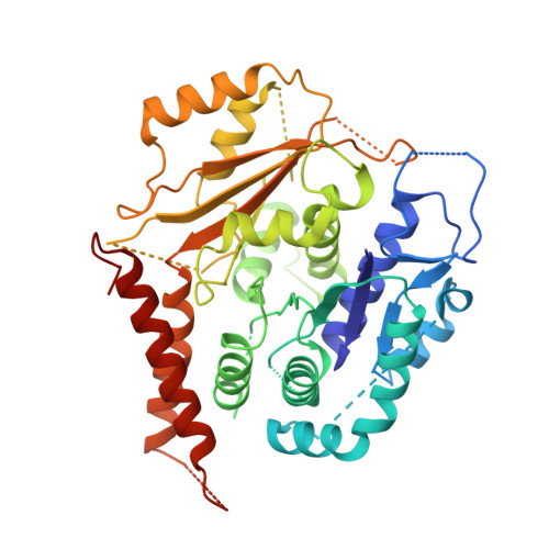
    The γ-tubulin ring complex (γ-TuRC) is an essential regulator of centrosomal and acentrosomal microtubule formation, yet its structure is not known. Here, we present a cryo-EM reconstruction of the native human γ-TuRC at ∼3.8 Å resolution, revealing an asymmetric, cone-shaped structure. Pseudo-atomic models indicate that GCP4, GCP5, and GCP6 form distinct Y-shaped assemblies that structurally mimic GCP2/GCP3 subcomplexes distal to the γ-TuRC "seam." We also identify an unanticipated structural bridge that includes an actin-like protein and spans the γ-TuRC lumen. Despite its asymmetric architecture, the γ-TuRC arranges γ-tubulins into a helical geometry poised to nucleate microtubules. Diversity in the γ-TuRC subunits introduces large (>100,000 Å 2 ) surfaces in the complex that allow for interactions with different regulatory factors. The observed compositional complexity of the γ-TuRC could self-regulate its assembly into a cone-shaped structure to control microtubule formation across diverse contexts, e.g., within biological condensates or alongside existing filaments.


  • Organizational Affiliation
    • Laboratory of Chemistry and Cell Biology, The Rockefeller University, 1230 York Avenue, New York, NY 10065, USA.

Macromolecule Content 

  • Total Structure Weight: 51.7 kDa 
  • Atom Count: 2,979 
  • Modeled Residue Count: 369 
  • Deposited Residue Count: 451 
  • Unique protein chains: 1

Macromolecules

Find similar proteins by:|  3D Structure
Entity ID: 1
MoleculeChains  Sequence LengthOrganismDetailsImage
Tubulin gamma-1 chainA [auth g]451Homo sapiensMutation(s): 0 
UniProt & NIH Common Fund Data Resources
Find proteins for P23258 (Homo sapiens)
Explore P23258 
Go to UniProtKB:  P23258
PHAROS:  P23258
GTEx:  ENSG00000131462 
Entity Groups
Sequence Clusters30% Identity50% Identity70% Identity90% Identity95% Identity100% Identity
UniProt GroupP23258
Sequence Annotations
Expand
Reference Sequence

Small Molecules

Ligands 1 Unique
IDChains Name / Formula / InChI Key2D Diagram3D Interactions
GDP

Query on GDP


Download:Ideal Coordinates CCD File
B [auth g]GUANOSINE-5'-DIPHOSPHATE
C10 H15 N5 O11 P2
QGWNDRXFNXRZMB-UUOKFMHZSA-N

Experimental Data & Validation

Experimental Data

  • Method: ELECTRON MICROSCOPY
  • Resolution: 3.80 Å
  • Aggregation State: PARTICLE 
  • Reconstruction Method: SINGLE PARTICLE 

Structure Validation

View Full Validation Report


Entry History 

& Funding Information

Deposition Data


Funding OrganizationLocationGrant Number
National Institutes of Health/National Institute of General Medical Sciences (NIH/NIGMS)United StatesR35 GM130234
Human Frontier Science Program (HFSP)FranceLT000025/18-L1

Revision History  (Full details and data files)

  • Version 1.0: 2020-01-01
    Type: Initial release
  • Version 1.1: 2020-01-22
    Changes: Database references
  • Version 1.2: 2024-03-06
    Changes: Data collection, Database references