6UX8

Structure of monobody 33 MLKL N-terminal domain complex


Experimental Data Snapshot

  • Method: X-RAY DIFFRACTION
  • Resolution: 2.50 Å
  • R-Value Free: 0.296 
  • R-Value Work: 0.247 
  • R-Value Observed: 0.249 

wwPDB Validation   3D Report Full Report

Currently 6UX8 does not have a validation slider image.


This is version 1.3 of the entry. See complete history


Literature

Identification of MLKL membrane translocation as a checkpoint in necroptotic cell death using Monobodies.

Petrie, E.J.Birkinshaw, R.W.Koide, A.Denbaum, E.Hildebrand, J.M.Garnish, S.E.Davies, K.A.Sandow, J.J.Samson, A.L.Gavin, X.Fitzgibbon, C.Young, S.N.Hennessy, P.J.Smith, P.P.C.Webb, A.I.Czabotar, P.E.Koide, S.Murphy, J.M.

(2020) Proc Natl Acad Sci U S A 117: 8468-8475

  • DOI: https://doi.org/10.1073/pnas.1919960117
  • Primary Citation of Related Structures:  
    6UX8

  • PubMed Abstract: 

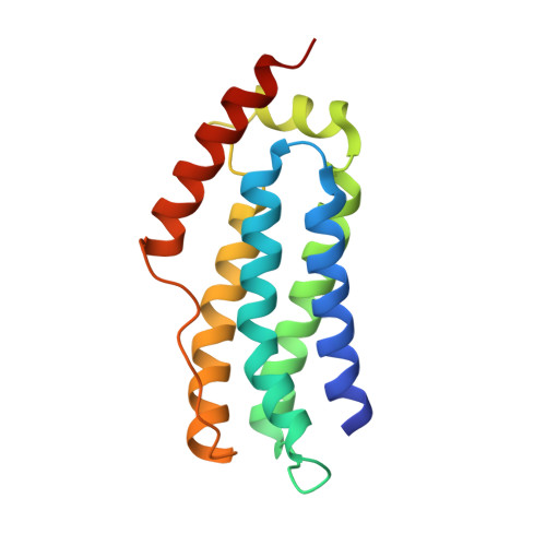
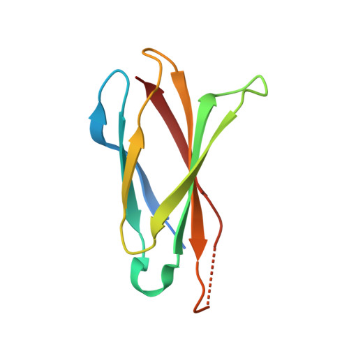
    The necroptosis cell death pathway has been implicated in host defense and in the pathology of inflammatory diseases. While phosphorylation of the necroptotic effector pseudokinase Mixed Lineage Kinase Domain-Like (MLKL) by the upstream protein kinase RIPK3 is a hallmark of pathway activation, the precise checkpoints in necroptosis signaling are still unclear. Here we have developed monobodies, synthetic binding proteins, that bind the N-terminal four-helix bundle (4HB) "killer" domain and neighboring first brace helix of human MLKL with nanomolar affinity. When expressed as genetically encoded reagents in cells, these monobodies potently block necroptotic cell death. However, they did not prevent MLKL recruitment to the "necrosome" and phosphorylation by RIPK3, nor the assembly of MLKL into oligomers, but did block MLKL translocation to membranes where activated MLKL normally disrupts membranes to kill cells. An X-ray crystal structure revealed a monobody-binding site centered on the α4 helix of the MLKL 4HB domain, which mutational analyses showed was crucial for reconstitution of necroptosis signaling. These data implicate the α4 helix of its 4HB domain as a crucial site for recruitment of adaptor proteins that mediate membrane translocation, distinct from known phospholipid binding sites.


  • Organizational Affiliation

    Walter and Eliza Hall Institute of Medical Research, Parkville, VIC 3052, Australia.


Macromolecules
Find similar proteins by:  (by identity cutoff)  |  3D Structure
Entity ID: 1
MoleculeChains Sequence LengthOrganismDetailsImage
Mixed lineage kinase domain-like protein158Homo sapiensMutation(s): 0 
Gene Names: MLKL
UniProt & NIH Common Fund Data Resources
Find proteins for Q8NB16 (Homo sapiens)
Explore Q8NB16 
Go to UniProtKB:  Q8NB16
PHAROS:  Q8NB16
GTEx:  ENSG00000168404 
Entity Groups  
Sequence Clusters30% Identity50% Identity70% Identity90% Identity95% Identity100% Identity
UniProt GroupQ8NB16
Sequence Annotations
Expand
  • Reference Sequence
Find similar proteins by:  (by identity cutoff)  |  3D Structure
Entity ID: 2
MoleculeChains Sequence LengthOrganismDetailsImage
Monobody97synthetic constructMutation(s): 0 
Gene Names: Monobody
Entity Groups  
Sequence Clusters30% Identity50% Identity70% Identity90% Identity95% Identity100% Identity
Sequence Annotations
Expand
  • Reference Sequence
Experimental Data & Validation

Experimental Data

  • Method: X-RAY DIFFRACTION
  • Resolution: 2.50 Å
  • R-Value Free: 0.296 
  • R-Value Work: 0.247 
  • R-Value Observed: 0.249 
  • Space Group: P 21 21 21
Unit Cell:
Length ( Å )Angle ( ˚ )
a = 58.294α = 90
b = 59.418β = 90
c = 68.043γ = 90
Software Package:
Software NamePurpose
PHENIXrefinement
XDSdata reduction
Aimlessdata scaling
PHASERphasing

Structure Validation

View Full Validation Report

Currently 6UX8 does not have a validation slider image.


Entry History 

Deposition Data

Revision History  (Full details and data files)

  • Version 1.0: 2020-04-01
    Type: Initial release
  • Version 1.1: 2020-04-15
    Changes: Database references
  • Version 1.2: 2020-04-29
    Changes: Database references
  • Version 1.3: 2023-10-11
    Changes: Data collection, Database references, Derived calculations, Refinement description