Biophysical characterization of two commercially available preparations of the drug containing Escherichia coli L-Asparaginase 2.
de Araujo, T.S., Scapin, S.M.N., de Andrade, W., Fasciotti, M., de Magalhaes, M.T.Q., Almeida, M.S., Lima, L.M.T.R.(2021) Biophys Chem 271: 106554-106554
- PubMed: 33607531 Search on PubMed
- DOI: https://doi.org/10.1016/j.bpc.2021.106554
- Primary Citation Related Structures:
6UOD, 6UOG, 6UOH - PubMed Abstract:
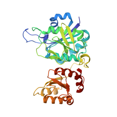
The hydrolysis of asparagine and glutamine by L-asparaginase has been used to treat acute lymphoblastic leukemia for over four decades. Each L-asparaginase monomer has a long loop that closes over the active site upon substrate binding, acting as a lid. Here we present a comparative study of two commercially available preparations of the drug containing Escherichia coli L-Asparaginase 2 (EcA2), performed by a comprehensive array of biophysical and biochemical approaches. We report the oligomeric landscape and conformational and dynamic plasticity of E. coli type 2 L-asparaginase present in two different formulations, and its relationship with L-aspartic acid, which is present in Aginasa, but not in Leuginase. The L-Asp present in Aginasa formulation was found to provide to EcA2 a resistance to in vitro proteolysis. EcA2 shows a composition of monomers and oligomers up to tetramers, which is mostly not altered in the presence of L-Asp. Ion-mobility spectrometry-mass spectrometry reveals two conformers for the monomeric EcA2, and that monomeric species has sufficient capacity for selective binding to L-Asp and L-Glu. The N-terminal loop of the EcA2 present in Leuginase, which is part of the active site is disordered, but it gets ordered in the presence of L-Asp, while L-Glu only does so to a limited extent. These data provide new insights on the mechanistic of ligand recognition by EcA2, and the impact of formulation in its conformational diversity landscape.
- Pharmaceutical Biotechnology Laboratory - pbiotech, Faculty of Pharmacy, Federal University of Rio de Janeiro, Rio de Janeiro, RJ 21941-902, Brazil; Protein Advanced Biochemistry - PAB, National Center for Structural Biology and Bioimaging - CENABIO, Federal University of Rio de Janeiro, Rio de Janeiro, RJ, 21941-902, Brazil; Institute for Medical Biochemistry Leopoldo DeMeis, Federal University of Rio de Janeiro, Rio de Janeiro, RJ, 21941-902, Brazil.
Organizational Affiliation: