6UHM

Crystal Structure of a Physical Mixture of C148 mGFP and scDNA-1


Experimental Data Snapshot

  • Method: X-RAY DIFFRACTION
  • Resolution: 2.10 Å
  • R-Value Free: 0.270 
  • R-Value Work: 0.211 
  • R-Value Observed: 0.214 

wwPDB Validation   3D Report Full Report


This is version 1.3 of the entry. See complete history


Literature

DNA-Directed Protein Packing within Single Crystals.

Winegar, P.H.Hayes, O.G.McMillan, J.R.Figg, C.A.Focia, P.J.Mirkin, C.A.

(2020) Chem 6: 1007-1017

  • DOI: https://doi.org/10.1016/j.chempr.2020.03.002
  • Primary Citation of Related Structures:  
    6UHJ, 6UHK, 6UHL, 6UHM, 6UHN, 6UHO, 6UHP, 6UHQ, 6UHR

  • PubMed Abstract: 

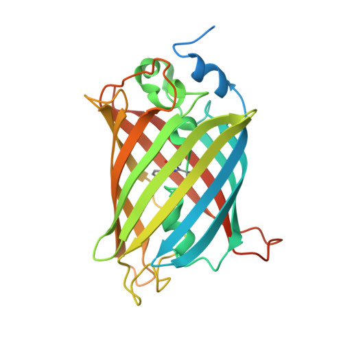
    Designed DNA-DNA interactions are investigated for their ability to modulate protein packing within single crystals of mutant green fluorescent proteins (mGFPs) functionalized with a single DNA strand (mGFP-DNA). We probe the effects of DNA sequence, length, and protein-attachment position on the formation and protein packing of mGFP-DNA crystals. Notably, when complementary mGFP-DNA conjugates are introduced to one another, crystals form with nearly identical packing parameters, regardless of sequence if the number of bases is equivalent. DNA complementarity is essential, because experiments with non-complementary sequences produce crystals with different protein arrangements. Importantly, the DNA length and its position of attachment on the protein markedly influence the formation of and protein packing within single crystals. This work shows how designed DNA interactions can be used to influence the growth and packing in X-ray diffraction quality protein single crystals and is thus an important step forward in protein crystal engineering.


  • Organizational Affiliation

    Department of Chemistry, Northwestern University, 2145 Sheridan Road, Evanston, IL 60208, USA.


Macromolecules
Find similar proteins by:  (by identity cutoff)  |  3D Structure
Entity ID: 1
MoleculeChains Sequence LengthOrganismDetailsImage
C148 mGFP
A, B
272Aequorea victoriaMutation(s): 0 
UniProt
Find proteins for P42212 (Aequorea victoria)
Explore P42212 
Go to UniProtKB:  P42212
Entity Groups  
Sequence Clusters30% Identity50% Identity70% Identity90% Identity95% Identity100% Identity
UniProt GroupP42212
Sequence Annotations
Expand
  • Reference Sequence
Small Molecules
Modified Residues  1 Unique
IDChains TypeFormula2D DiagramParent
CRO
Query on CRO
A, B
L-PEPTIDE LINKINGC15 H17 N3 O5THR, TYR, GLY
Experimental Data & Validation

Experimental Data

  • Method: X-RAY DIFFRACTION
  • Resolution: 2.10 Å
  • R-Value Free: 0.270 
  • R-Value Work: 0.211 
  • R-Value Observed: 0.214 
  • Space Group: P 21 21 21
Unit Cell:
Length ( Å )Angle ( ˚ )
a = 58.28α = 90
b = 61.76β = 90
c = 135.32γ = 90
Software Package:
Software NamePurpose
REFMACrefinement
iMOSFLMdata reduction
SCALAdata scaling
PHASERphasing

Structure Validation

View Full Validation Report


Entry History & Funding Information

Deposition Data


Funding OrganizationLocationGrant Number
Department of Defense (DOD, United States)United StatesN00014-15-1-0043
Department of Defense (DOD, United States)United StatesFA9550-17-1-0348

Revision History  (Full details and data files)

  • Version 1.0: 2020-03-18
    Type: Initial release
  • Version 1.1: 2021-03-24
    Changes: Database references
  • Version 1.2: 2023-10-11
    Changes: Data collection, Database references, Refinement description
  • Version 1.3: 2023-11-15
    Changes: Data collection