The alpha-synuclein hereditary mutation E46K unlocks a more stable, pathogenic fibril structure.
Boyer, D.R., Li, B., Sun, C., Fan, W., Zhou, K., Hughes, M.P., Sawaya, M.R., Jiang, L., Eisenberg, D.S.(2020) Proc Natl Acad Sci U S A 117: 3592-3602
- PubMed: 32015135 Search on PubMedSearch on PubMed Central
- DOI: https://doi.org/10.1073/pnas.1917914117
- Primary Citation Related Structures:
6UFR - PubMed Abstract:
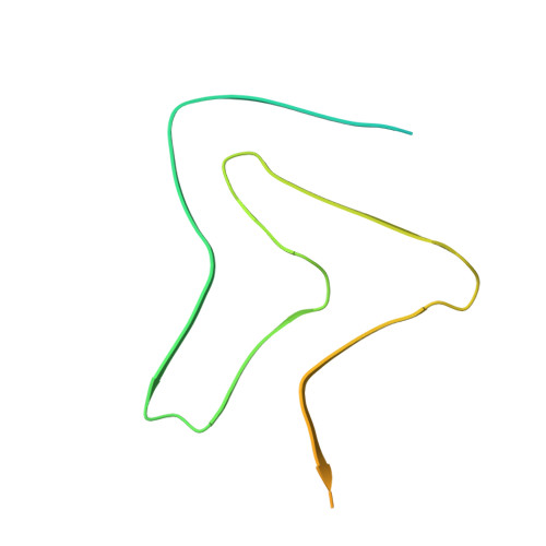
Aggregation of α-synuclein is a defining molecular feature of Parkinson's disease, Lewy body dementia, and multiple systems atrophy. Hereditary mutations in α-synuclein are linked to both Parkinson's disease and Lewy body dementia; in particular, patients bearing the E46K disease mutation manifest a clinical picture of parkinsonism and Lewy body dementia, and E46K creates more pathogenic fibrils in vitro. Understanding the effect of these hereditary mutations on α-synuclein fibril structure is fundamental to α-synuclein biology. We therefore determined the cryo-electron microscopy (cryo-EM) structure of α-synuclein fibrils containing the hereditary E46K mutation. The 2.5-Å structure reveals a symmetric double protofilament in which the molecules adopt a vastly rearranged, lower energy fold compared to wild-type fibrils. We propose that the E46K misfolding pathway avoids electrostatic repulsion between K46 and K80, a residue pair which form the E46-K80 salt bridge in the wild-type fibril structure. We hypothesize that, under our conditions, the wild-type fold does not reach this deeper energy well of the E46K fold because the E46-K80 salt bridge diverts α-synuclein into a kinetic trap-a shallower, more accessible energy minimum. The E46K mutation apparently unlocks a more stable and pathogenic fibril structure.
- Department of Chemistry and Biochemistry, University of California, Los Angeles, CA 90095.
Organizational Affiliation: