6UEK

Structure of Urocanate Hydratase from Trypanosoma cruzi in complex with NAD+


Experimental Data Snapshot

  • Method: X-RAY DIFFRACTION
  • Resolution: 2.16 Å
  • R-Value Free: 0.255 
  • R-Value Work: 0.218 

wwPDB Validation   3D Report Full Report


Ligand Structure Quality Assessment 


This is version 1.2 of the entry. See complete history


Literature

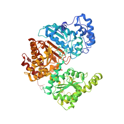
Structure of Urocanate Hydratase from the protozoan Trypanosoma cruzi.

Boreiko, S.Silva, M.de F P Melo, R.Silber, A.M.Iulek, J.

(2019) Int J Biol Macromol 146: 716-724

  • DOI: https://doi.org/10.1016/j.ijbiomac.2019.12.101
  • Primary Citation of Related Structures:  
    6UEK

  • PubMed Abstract: 

    The enzyme Urocanate Hydratase (UH) participates in the catabolic pathway of L-histidine. Trypanosoma cruzi Urocanate Hydratase (TcUH) is identified as a therapeutic molecular target in the WHO/TDR Targets Database. We report the 3D structure determination and number of features of TcUH, and compared it to other few available bacterial UH structures. Each monomer presents two domains and one NAD + molecule. Superpositions revealed differences in the relative orientation of domains within monomers, such that TcUH monomer A resembles Urocanate Hydratase from Geobacillus kaustophilus (GkUH) (open conformation), while monomer C resembles Urocanate Hydratase from Pseudomonas putida (PpUH) and Urocanate Hydratase from Bacillus subtilis (BsUH) (closed conformations). We use the structure of TcUH to make considerations about 3 non-deleterious and 2 deleterious mutations found in human UHs: non-deleterious mutations could be accommodated without large displacements or interaction interruptions, whereas deleterious mutations in one case might disrupt an α-helix (as previously suggested) and in the other case, besides disrupting the enzyme interaction with the substrate, might interfere with interdomain movement.


  • Organizational Affiliation

    Department of Chemistry, State University of Ponta Grossa, Ponta Grossa, PR 84030-900, Brazil.


Macromolecules
Find similar proteins by:  (by identity cutoff)  |  3D Structure
Entity ID: 1
MoleculeChains Sequence LengthOrganismDetailsImage
Urocanate hydratase
A, B, C, D
681Trypanosoma cruziMutation(s): 0 
UniProt
Find proteins for Q4D9S6 (Trypanosoma cruzi (strain CL Brener))
Explore Q4D9S6 
Go to UniProtKB:  Q4D9S6
Entity Groups  
Sequence Clusters30% Identity50% Identity70% Identity90% Identity95% Identity100% Identity
UniProt GroupQ4D9S6
Sequence Annotations
Expand
  • Reference Sequence
Experimental Data & Validation

Experimental Data

  • Method: X-RAY DIFFRACTION
  • Resolution: 2.16 Å
  • R-Value Free: 0.255 
  • R-Value Work: 0.218 
  • Space Group: P 1 21 1
Unit Cell:
Length ( Å )Angle ( ˚ )
a = 84.614α = 90
b = 134.788β = 93.25
c = 113.399γ = 90
Software Package:
Software NamePurpose
PHENIXrefinement
PDB_EXTRACTdata extraction
XDSdata reduction
XDSdata scaling
PHASERphasing

Structure Validation

View Full Validation Report


Ligand Structure Quality Assessment 


Entry History & Funding Information

Deposition Data


Funding OrganizationLocationGrant Number
Brazilian National Council for Scientific and Technological Development (CNPq)Brazil573607/2008-7 and 08/57910-0

Revision History  (Full details and data files)

  • Version 1.0: 2020-01-15
    Type: Initial release
  • Version 1.1: 2020-02-05
    Changes: Database references
  • Version 1.2: 2023-10-11
    Changes: Data collection, Database references, Refinement description