6U7L

2.75 Angstrom Crystal Structure of Galactarate Dehydratase from Escherichia coli.


Experimental Data Snapshot

  • Method: X-RAY DIFFRACTION
  • Resolution: 2.75 Å
  • R-Value Free: 0.241 
  • R-Value Work: 0.206 
  • R-Value Observed: 0.208 

wwPDB Validation   3D Report Full Report


This is version 1.1 of the entry. See complete history


Literature

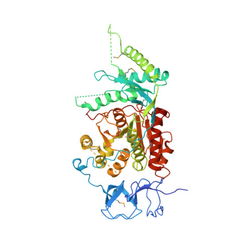
Structure of galactarate dehydratase, a new fold in an enolase involved in bacterial fitness after antibiotic treatment.

Rosas-Lemus, M.Minasov, G.Shuvalova, L.Wawrzak, Z.Kiryukhina, O.Mih, N.Jaroszewski, L.Palsson, B.Godzik, A.Satchell, K.J.F.

(2020) Protein Sci 29: 711-722

  • DOI: 10.1002/pro.3796
  • Primary Citation of Related Structures:  
    6U7L

  • PubMed Abstract: 
  • Galactarate dehydratase (GarD) is the first enzyme in the galactarate/glucarate pathway and catalyzes the dehydration of galactarate to 3-keto-5-dehydroxygalactarate. This protein is known to increase colonization fitness of intestinal pathogens in antibiotic-treated mice and to promote bacterial survival during stress ...

    Galactarate dehydratase (GarD) is the first enzyme in the galactarate/glucarate pathway and catalyzes the dehydration of galactarate to 3-keto-5-dehydroxygalactarate. This protein is known to increase colonization fitness of intestinal pathogens in antibiotic-treated mice and to promote bacterial survival during stress. The galactarate/glucarate pathway is widespread in bacteria, but not in humans, and thus could be a target to develop new inhibitors for use in combination therapy to combat antibiotic resistance. The structure of almost all the enzymes of the galactarate/glucarate pathway were solved previously, except for GarD, for which only the structure of the N-terminal domain was determined previously. Herein, we report the first crystal structure of full-length GarD solved using a seleno-methoionine derivative revealing a new protein fold. The protein consists of three domains, each presenting a novel twist as compared to their distant homologs. GarD in the crystal structure forms dimers and each monomer consists of three domains. The N-terminal domain is comprised of a β-clip fold, connected to the second domain by a long unstructured linker. The second domain serves as a dimerization interface between two monomers. The C-terminal domain forms an unusual variant of a Rossmann fold with a crossover and is built around a seven-stranded parallel β-sheet supported by nine α-helices. A metal binding site in the C-terminal domain is occupied by Ca 2+ . The activity of GarD was corroborated by the production of 5-keto-4-deoxy-D-glucarate under reducing conditions and in the presence of iron. Thus, GarD is an unusual enolase with a novel protein fold never previously seen in this class of enzymes.


    Organizational Affiliation

    Center for Structural Genomics of Infectious Diseases, Northwestern University, Feinberg School of Medicine, Chicago, Illinois.


Macromolecules
Find similar proteins by:  (by identity cutoff)  |  3D Structure
Entity ID: 1
MoleculeChainsSequence LengthOrganismDetailsImage
Galactarate dehydratase (L-threo-forming)A, B526Escherichia coli K-12Mutation(s): 0 
Gene Names: garDyhaGb3128JW3097
EC: 4.2.1.42
UniProt
Find proteins for P39829 (Escherichia coli (strain K12))
Explore P39829 
Go to UniProtKB:  P39829
Entity Groups  
Sequence Clusters30% Identity50% Identity70% Identity90% Identity95% Identity100% Identity
UniProt GroupP39829
Protein Feature View
Expand
  • Reference Sequence
Find similar proteins by:  (by identity cutoff)  |  3D Structure
Entity ID: 2
MoleculeChainsSequence LengthOrganismDetailsImage
Galactarate dehydratase (L-threo-forming)C, D526Escherichia coli K-12Mutation(s): 0 
Gene Names: garDyhaGb3128JW3097
EC: 4.2.1.42
UniProt
Find proteins for P39829 (Escherichia coli (strain K12))
Explore P39829 
Go to UniProtKB:  P39829
Entity Groups  
Sequence Clusters30% Identity50% Identity70% Identity90% Identity95% Identity100% Identity
UniProt GroupP39829
Protein Feature View
Expand
  • Reference Sequence
Small Molecules
Ligands 2 Unique
IDChainsName / Formula / InChI Key2D Diagram3D Interactions
CA (Subject of Investigation/LOI)
Query on CA

Download Ideal Coordinates CCD File 
E [auth A],
F [auth A],
G [auth A],
L [auth B],
M [auth B],
E [auth A],
F [auth A],
G [auth A],
L [auth B],
M [auth B],
N [auth C],
O [auth C],
P [auth D],
Q [auth D]
CALCIUM ION
Ca
BHPQYMZQTOCNFJ-UHFFFAOYSA-N
 Ligand Interaction
CL
Query on CL

Download Ideal Coordinates CCD File 
H [auth A],
I [auth A],
J [auth A],
K [auth A],
R [auth D],
H [auth A],
I [auth A],
J [auth A],
K [auth A],
R [auth D],
S [auth D],
T [auth D]
CHLORIDE ION
Cl
VEXZGXHMUGYJMC-UHFFFAOYSA-M
 Ligand Interaction
Modified Residues  2 Unique
IDChainsTypeFormula2D DiagramParent
MSE
Query on MSE
A, B L-PEPTIDE LINKINGC5 H11 N O2 SeMET
PHD
Query on PHD
A, B L-PEPTIDE LINKINGC4 H8 N O7 PASP
Experimental Data & Validation

Experimental Data

  • Method: X-RAY DIFFRACTION
  • Resolution: 2.75 Å
  • R-Value Free: 0.241 
  • R-Value Work: 0.206 
  • R-Value Observed: 0.208 
  • Space Group: P 1 21 1
Unit Cell:
Length ( Å )Angle ( ˚ )
a = 58.462α = 90
b = 167.574β = 103.97
c = 117.065γ = 90
Software Package:
Software NamePurpose
REFMACrefinement
PDB_EXTRACTdata extraction
HKL-3000data reduction
HKL-3000data scaling
PHENIXphasing

Structure Validation

View Full Validation Report


Entry History 

Revision History  (Full details and data files)

  • Version 1.0: 2019-11-06
    Type: Initial release
  • Version 1.1: 2021-01-27
    Changes: Database references, Derived calculations