6U3W | pdb_00006u3w

Crystal structure of yeast alpha/epsilon-COP of the COPI vesicular coat


Experimental Data Snapshot

  • Method: X-RAY DIFFRACTION
  • Resolution: 2.39 Å
  • R-Value Free: 
    0.238 (Depositor), 0.237 (DCC) 
  • R-Value Work: 
    0.178 (Depositor), 0.179 (DCC) 
  • R-Value Observed: 
    0.182 (Depositor) 

Starting Model: experimental
View more details

wwPDB Validation 3D Report Full Report

Validation slider image for 6U3W

This is version 1.4 of the entry. See complete history

Literature

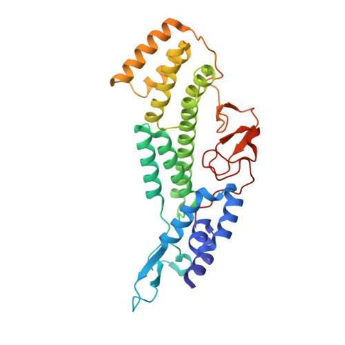
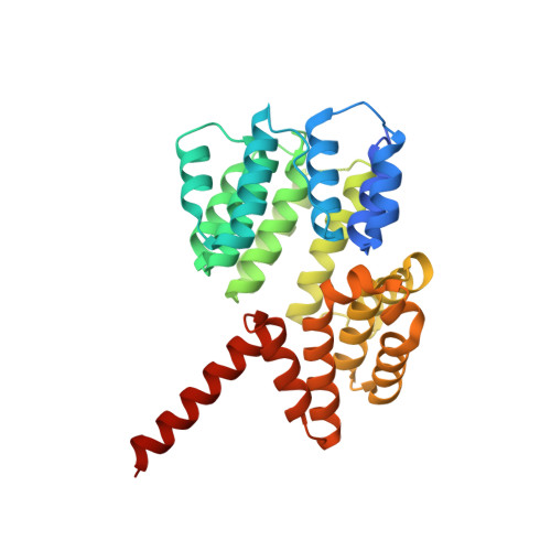
Roles of singleton tryptophan motifs in COPI coat stability and vesicle tethering.

Travis, S.M.Kokona, B.Fairman, R.Hughson, F.M.

(2019) Proc Natl Acad Sci U S A 116: 24031-24040

  • DOI: https://doi.org/10.1073/pnas.1909697116
  • Primary Citation Related Structures: 
    6TZT, 6U3V, 6U3W

  • PubMed Abstract: 

    Coat protein I (COPI)-coated vesicles mediate retrograde transport from the Golgi to the endoplasmic reticulum (ER), as well as transport within the Golgi. Major progress has been made in defining the structure of COPI coats, in vitro and in vivo, at resolutions as high as 9 Å. Nevertheless, important questions remain unanswered, including what specific interactions stabilize COPI coats, how COPI vesicles recognize their target membranes, and how coat disassembly is coordinated with vesicle fusion and cargo delivery. Here, we use X-ray crystallography to identify a conserved site on the COPI subunit α-COP that binds to flexible, acidic sequences containing a single tryptophan residue. One such sequence, found within α-COP itself, mediates α-COP homo-oligomerization. Another such sequence is contained within the lasso of the ER-resident Dsl1 complex, where it helps mediate the tethering of Golgi-derived COPI vesicles at the ER membrane. Together, our findings suggest that α-COP homo-oligomerization plays a key role in COPI coat stability, with potential implications for the coordination of vesicle tethering, uncoating, and fusion.


  • Organizational Affiliation
    • Department of Molecular Biology, Princeton University, Princeton, NJ 08544.

Macromolecule Content 

  • Total Structure Weight: 69.55 kDa 
  • Atom Count: 4,915 
  • Modeled Residue Count: 594 
  • Deposited Residue Count: 613 
  • Unique protein chains: 2

Macromolecules

Find similar proteins by:|  3D Structure
Entity ID: 1
MoleculeChains  Sequence LengthOrganismDetailsImage
Coatomer subunit alpha303Saccharomyces cerevisiae S288CMutation(s): 0 
Gene Names: COP1RET1SEC33SOO1YDL145CD1578
UniProt
Find proteins for P53622 (Saccharomyces cerevisiae (strain ATCC 204508 / S288c))
Explore P53622 
Go to UniProtKB:  P53622
Entity Groups
Sequence Clusters30% Identity50% Identity70% Identity90% Identity95% Identity100% Identity
UniProt GroupP53622
Sequence Annotations
Expand
Reference Sequence
Find similar proteins by:|  3D Structure
Entity ID: 2
MoleculeChains  Sequence LengthOrganismDetailsImage
Coatomer subunit epsilon310Saccharomyces cerevisiae S288CMutation(s): 0 
Gene Names: SEC28YIL076W
UniProt
Find proteins for P40509 (Saccharomyces cerevisiae (strain ATCC 204508 / S288c))
Explore P40509 
Go to UniProtKB:  P40509
Entity Groups
Sequence Clusters30% Identity50% Identity70% Identity90% Identity95% Identity100% Identity
UniProt GroupP40509
Sequence Annotations
Expand
Reference Sequence

Small Molecules

Ligands 1 Unique
IDChains Name / Formula / InChI Key2D Diagram3D Interactions
GOL

Query on GOL


Download:Ideal Coordinates CCD File
C [auth A]GLYCEROL
C3 H8 O3
PEDCQBHIVMGVHV-UHFFFAOYSA-N

Experimental Data & Validation

Experimental Data

  • Method: X-RAY DIFFRACTION
  • Resolution: 2.39 Å
  • R-Value Free:  0.238 (Depositor), 0.237 (DCC) 
  • R-Value Work:  0.178 (Depositor), 0.179 (DCC) 
  • R-Value Observed: 0.182 (Depositor) 
Space Group: P 21 21 21
Unit Cell:
Length ( Å )Angle ( ˚ )
a = 72.264α = 90
b = 84.594β = 90
c = 117.842γ = 90
Software Package:
Software NamePurpose
DENZOdata reduction
SCALEPACKdata scaling
PHASERphasing
PHENIXrefinement
PDB_EXTRACTdata extraction

Structure Validation

View Full Validation Report


Entry History 

& Funding Information

Deposition Data


Funding OrganizationLocationGrant Number
National Institutes of Health/National Institute of General Medical Sciences (NIH/NIGMS)United StatesR01GM071574
National Institutes of Health/National Institute of General Medical Sciences (NIH/NIGMS)United StatesF31GM12676

Revision History  (Full details and data files)

  • Version 1.0: 2019-11-13
    Type: Initial release
  • Version 1.1: 2019-11-27
    Changes: Database references
  • Version 1.2: 2019-12-11
    Changes: Database references
  • Version 1.3: 2020-01-01
    Changes: Author supporting evidence
  • Version 1.4: 2023-10-11
    Changes: Data collection, Database references, Refinement description