6U0T

Protofilament Ribbon Flagellar Proteins Rib43a-S


Experimental Data Snapshot

  • Method: ELECTRON MICROSCOPY
  • Resolution: 4.16 Å
  • Aggregation State: FILAMENT 
  • Reconstruction Method: SINGLE PARTICLE 

wwPDB Validation   3D Report Full Report


This is version 1.4 of the entry. See complete history


Literature

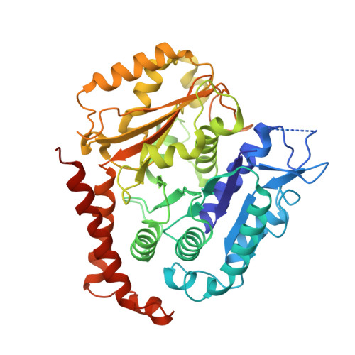
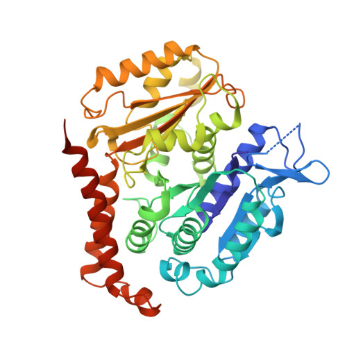
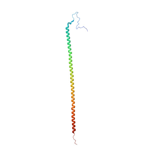
Tubulin lattice in cilia is in a stressed form regulated by microtubule inner proteins.

Ichikawa, M.Khalifa, A.A.Z.Kubo, S.Dai, D.Basu, K.Maghrebi, M.A.F.Vargas, J.Bui, K.H.

(2019) Proc Natl Acad Sci U S A 116: 19930-19938

  • DOI: https://doi.org/10.1073/pnas.1911119116
  • Primary Citation of Related Structures:  
    6U0H, 6U0T, 6U0U

  • PubMed Abstract: 

    Cilia, the hair-like protrusions that beat at high frequencies to propel a cell or move fluid around are composed of radially bundled doublet microtubules. In this study, we present a near-atomic resolution map of the Tetrahymena doublet microtubule by cryoelectron microscopy. The map demonstrates that the network of microtubule inner proteins weaves into the tubulin lattice and forms an inner sheath. From mass spectrometry data and de novo modeling, we identified Rib43a proteins as the filamentous microtubule inner proteins in the protofilament ribbon region. The Rib43a-tubulin interaction leads to an elongated tubulin dimer distance every 2 dimers. In addition, the tubulin lattice structure with missing microtubule inner proteins (MIPs) by sarkosyl treatment shows significant longitudinal compaction and lateral angle change between protofilaments. These results are evidence that the MIPs directly affect and stabilize the tubulin lattice. It suggests that the doublet microtubule is an intrinsically stressed filament and that this stress could be manipulated in the regulation of ciliary waveforms.


  • Organizational Affiliation

    Department of Anatomy and Cell Biology, McGill University, Montréal, QC H3A 0C7, Canada.


Macromolecules
Find similar proteins by:  (by identity cutoff)  |  3D Structure
Entity ID: 1
MoleculeChains Sequence LengthOrganismDetailsImage
Tubulin alpha chain449Tetrahymena thermophilaMutation(s): 0 
UniProt
Find proteins for P41351 (Tetrahymena thermophila)
Explore P41351 
Go to UniProtKB:  P41351
Entity Groups  
Sequence Clusters30% Identity50% Identity70% Identity90% Identity95% Identity100% Identity
UniProt GroupP41351
Sequence Annotations
Expand
  • Reference Sequence
Find similar proteins by:  (by identity cutoff)  |  3D Structure
Entity ID: 2
MoleculeChains Sequence LengthOrganismDetailsImage
Tubulin beta chain443Tetrahymena thermophilaMutation(s): 0 
Gene Names: BTU1BTU2
UniProt
Find proteins for P41352 (Tetrahymena thermophila)
Explore P41352 
Go to UniProtKB:  P41352
Entity Groups  
Sequence Clusters30% Identity50% Identity70% Identity90% Identity95% Identity100% Identity
UniProt GroupP41352
Sequence Annotations
Expand
  • Reference Sequence
Find similar proteins by:  (by identity cutoff)  |  3D Structure
Entity ID: 3
MoleculeChains Sequence LengthOrganismDetailsImage
RIB43A proteinM [auth A]142Tetrahymena thermophila SB210Mutation(s): 0 
Gene Names: TTHERM_00641119
UniProt
Find proteins for A4VDZ5 (Tetrahymena thermophila (strain SB210))
Explore A4VDZ5 
Go to UniProtKB:  A4VDZ5
Entity Groups  
Sequence Clusters30% Identity50% Identity70% Identity90% Identity95% Identity100% Identity
UniProt GroupA4VDZ5
Sequence Annotations
Expand
  • Reference Sequence
Small Molecules
Ligands 3 Unique
IDChains Name / Formula / InChI Key2D Diagram3D Interactions
GTP
Query on GTP

Download Ideal Coordinates CCD File 
CA [auth H]
N [auth C]
Q [auth D]
T [auth E]
W [auth F]
CA [auth H],
N [auth C],
Q [auth D],
T [auth E],
W [auth F],
Z [auth G]
GUANOSINE-5'-TRIPHOSPHATE
C10 H16 N5 O14 P3
XKMLYUALXHKNFT-UUOKFMHZSA-N
GDP
Query on GDP

Download Ideal Coordinates CCD File 
BA [auth L]
EA [auth M]
P [auth B]
S [auth I]
V [auth J]
BA [auth L],
EA [auth M],
P [auth B],
S [auth I],
V [auth J],
Y [auth K]
GUANOSINE-5'-DIPHOSPHATE
C10 H15 N5 O11 P2
QGWNDRXFNXRZMB-UUOKFMHZSA-N
MG
Query on MG

Download Ideal Coordinates CCD File 
AA [auth G]
DA [auth H]
O [auth C]
R [auth D]
U [auth E]
AA [auth G],
DA [auth H],
O [auth C],
R [auth D],
U [auth E],
X [auth F]
MAGNESIUM ION
Mg
JLVVSXFLKOJNIY-UHFFFAOYSA-N
Experimental Data & Validation

Experimental Data

  • Method: ELECTRON MICROSCOPY
  • Resolution: 4.16 Å
  • Aggregation State: FILAMENT 
  • Reconstruction Method: SINGLE PARTICLE 
EM Software:
TaskSoftware PackageVersion
MODEL REFINEMENTPHENIX1.14
MODEL REFINEMENTCoot0.8.7
RECONSTRUCTIONRELION3.0

Structure Validation

View Full Validation Report


Entry History & Funding Information

Deposition Data


Funding OrganizationLocationGrant Number
Natural Sciences and Engineering Research Council (NSERC, Canada)CanadaDG69462
Canadian Institutes of Health Research (CIHR)CanadaPG388933
Natural Sciences and Engineering Research Council (NSERC, Canada)CanadaRGPIN-2018-04813

Revision History  (Full details and data files)

  • Version 1.0: 2019-09-25
    Type: Initial release
  • Version 1.1: 2019-10-02
    Changes: Data collection, Database references
  • Version 1.2: 2019-10-16
    Changes: Data collection, Database references
  • Version 1.3: 2020-01-08
    Changes: Author supporting evidence
  • Version 1.4: 2024-03-13
    Changes: Data collection, Database references, Derived calculations