6TV6 | pdb_00006tv6

Octameric McsB from Bacillus subtilis.


Experimental Data Snapshot

  • Method: X-RAY DIFFRACTION
  • Resolution: 2.50 Å
  • R-Value Free: 
    0.237 (Depositor), 0.241 (DCC) 
  • R-Value Work: 
    0.195 (Depositor), 0.199 (DCC) 
  • R-Value Observed: 
    0.195 (Depositor) 

Starting Model: experimental
View more details

wwPDB Validation 3D Report Full Report

Validation slider image for 6TV6

This is version 1.2 of the entry. See complete history

Literature

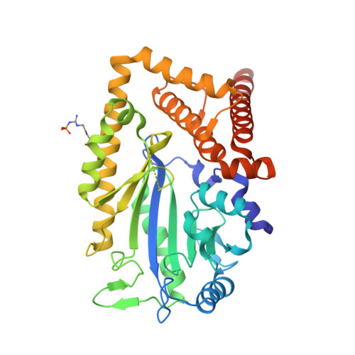
McsB forms a gated kinase chamber to mark aberrant bacterial proteins for degradation.

Hajdusits, B.Suskiewicz, M.J.Hundt, N.Meinhart, A.Kurzbauer, R.Leodolter, J.Kukura, P.Clausen, T.

(2021) Elife 10

  • DOI: https://doi.org/10.7554/eLife.63505
  • Primary Citation Related Structures: 
    6TV6

  • PubMed Abstract: 

    In Gram-positive bacteria, the McsB protein arginine kinase is central to protein quality control, labeling aberrant molecules for degradation by the ClpCP protease. Despite its importance for stress response and pathogenicity, it is still elusive how the bacterial degradation labeling is regulated. Here, we delineate the mechanism how McsB targets aberrant proteins during stress conditions. Structural data reveal a self-compartmentalized kinase, in which the active sites are sequestered in a molecular cage. The 'closed' octamer interconverts with other oligomers in a phosphorylation-dependent manner and, unlike these 'open' forms, preferentially labels unfolded proteins. In vivo data show that heat-shock triggers accumulation of higher order oligomers, of which the octameric McsB is essential for surviving stress situations. The interconversion of open and closed oligomers represents a distinct regulatory mechanism of a degradation labeler, allowing the McsB kinase to adapt its potentially dangerous enzyme function to the needs of the bacterial cell.


  • Organizational Affiliation
    • Research Institute of Molecular Pathology (IMP), Vienna BioCenter, Vienna, Austria.

Macromolecule Content 

  • Total Structure Weight: 170.53 kDa 
  • Atom Count: 11,404 
  • Modeled Residue Count: 1,418 
  • Deposited Residue Count: 1,496 
  • Unique protein chains: 1

Macromolecules

Find similar proteins by:|  3D Structure
Entity ID: 1
MoleculeChains  Sequence LengthOrganismDetailsImage
Protein-arginine kinase
A, B, C, D
374Bacillus subtilis subsp. subtilis str. 168Mutation(s): 0 
Gene Names: mcsByacIBSU00850
EC: 2.7.14.1
UniProt
Find proteins for P37570 (Bacillus subtilis (strain 168))
Explore P37570 
Go to UniProtKB:  P37570
Entity Groups
Sequence Clusters30% Identity50% Identity70% Identity90% Identity95% Identity100% Identity
UniProt GroupP37570
Sequence Annotations
Expand
Reference Sequence

Experimental Data & Validation

Experimental Data

  • Method: X-RAY DIFFRACTION
  • Resolution: 2.50 Å
  • R-Value Free:  0.237 (Depositor), 0.241 (DCC) 
  • R-Value Work:  0.195 (Depositor), 0.199 (DCC) 
  • R-Value Observed: 0.195 (Depositor) 
Space Group: P 21 21 2
Unit Cell:
Length ( Å )Angle ( ˚ )
a = 137.79α = 90
b = 147.64β = 90
c = 81.47γ = 90
Software Package:
Software NamePurpose
XDSdata reduction
XSCALEdata scaling
PHASERphasing
PHENIXrefinement
Cootmodel building

Structure Validation

View Full Validation Report


Entry History 

& Funding Information

Deposition Data


Funding OrganizationLocationGrant Number
European Research Council (ERC)Austria694978

Revision History  (Full details and data files)

  • Version 1.0: 2021-07-21
    Type: Initial release
  • Version 1.1: 2022-08-03
    Changes: Database references, Derived calculations
  • Version 1.2: 2024-01-24
    Changes: Data collection, Derived calculations, Refinement description