6TJP

Crystal structure of T7 bacteriophage portal protein, 13mer, closed valve - P212121


Experimental Data Snapshot

  • Method: X-RAY DIFFRACTION
  • Resolution: 3.74 Å
  • R-Value Free: 0.286 
  • R-Value Work: 0.244 
  • R-Value Observed: 0.246 

wwPDB Validation   3D Report Full Report


This is version 1.2 of the entry. See complete history


Literature

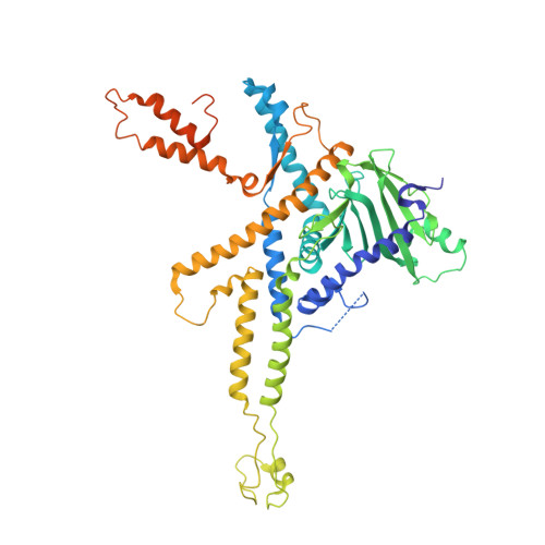
Using a partial atomic model from medium-resolution cryo-EM to solve a large crystal structure.

Fabrega-Ferrer, M.Cuervo, A.Fernandez, F.J.Machon, C.Perez-Luque, R.Pous, J.Vega, M.C.Carrascosa, J.L.Coll, M.

(2021) Acta Crystallogr D Struct Biol 77: 11-18

  • DOI: https://doi.org/10.1107/S2059798320015156
  • Primary Citation of Related Structures:  
    6TJP

  • PubMed Abstract: 

    Medium-resolution cryo-electron microscopy maps, in particular when they include a significant number of α-helices, may allow the building of partial models that are useful for molecular-replacement searches in large crystallographic structures when the structures of homologs are not available and experimental phasing has failed. Here, as an example, the solution of the structure of a bacteriophage portal using a partial 30% model built into a 7.8 Å resolution cryo-EM map is shown. Inspection of the self-rotation function allowed the correct oligomerization state to be determined, and density-modification procedures using rotation matrices and a mask based on the cryo-EM structure were critical for solving the structure. A workflow is described that may be applicable to similar cases and this strategy is compared with direct use of the cryo-EM map for molecular replacement.


  • Organizational Affiliation

    Institute for Research in Biomedicine (IRB Barcelona), The Barcelona Institute of Science and Technology, Baldiri Reixac 10, 08028 Barcelona, Spain.


Macromolecules
Find similar proteins by:  (by identity cutoff)  |  3D Structure
Entity ID: 1
MoleculeChains Sequence LengthOrganismDetailsImage
Portal protein
A, B, C, D, E
A, B, C, D, E, F, G, H, I, J, K, L, M
536Escherichia phage T7Mutation(s): 0 
UniProt
Find proteins for P03728 (Escherichia phage T7)
Explore P03728 
Go to UniProtKB:  P03728
Entity Groups  
Sequence Clusters30% Identity50% Identity70% Identity90% Identity95% Identity100% Identity
UniProt GroupP03728
Sequence Annotations
Expand
  • Reference Sequence
Experimental Data & Validation

Experimental Data

  • Method: X-RAY DIFFRACTION
  • Resolution: 3.74 Å
  • R-Value Free: 0.286 
  • R-Value Work: 0.244 
  • R-Value Observed: 0.246 
  • Space Group: P 21 21 21
Unit Cell:
Length ( Å )Angle ( ˚ )
a = 119.85α = 90
b = 238.57β = 90
c = 265.61γ = 90
Software Package:
Software NamePurpose
REFMACrefinement
PHENIXrefinement
XDSdata reduction
XSCALEdata scaling
PHASERphasing

Structure Validation

View Full Validation Report


Entry History & Funding Information

Deposition Data


Funding OrganizationLocationGrant Number
Spanish Ministry of Science, Innovation, and UniversitiesSpainBFU 2014-54181
Spanish Ministry of Science, Innovation, and UniversitiesSpainBFU2014-53550-P
Spanish Ministry of Science, Innovation, and UniversitiesSpainBFU2017-83720-P
Spanish Ministry of Science, Innovation, and UniversitiesSpainMDM-2014-0435
Spanish Ministry of Science, Innovation, and UniversitiesSpainSEV-2013-0347
Spanish Ministry of Science, Innovation, and UniversitiesSpainRYC-2011-09071
European Commission653706

Revision History  (Full details and data files)

  • Version 1.0: 2020-12-16
    Type: Initial release
  • Version 1.1: 2021-06-30
    Changes: Database references
  • Version 1.2: 2024-05-01
    Changes: Advisory, Data collection, Database references, Refinement description