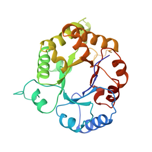
The adaptability of the active site of trypanosomal triosephosphate isomerase as observed in the crystal structures of three different complexes.
Noble, M.E., Wierenga, R.K., Lambeir, A.M., Opperdoes, F.R., Thunnissen, A.M., Kalk, K.H., Groendijk, H., Hol, W.G.(1991) Proteins 10: 50-69
- PubMed: 2062828 Search on PubMed
- DOI: https://doi.org/10.1002/prot.340100106
- Primary Citation Related Structures:
1IIG, 1IIH, 6TIM - PubMed Abstract:
Crystals of triosephosphate isomerase from Trypanosoma brucei brucei have been used in binding studies with three competitive inhibitors of the enzyme's activity. Highly refined structures have been deduced for the complexes between trypanosomal triosephosphate isomerase and a substrate analogue (glycerol-3-phosphate to 2.2 A), a transition state analogue (3-phosphonopropionic acid to 2.6 A), and a compound structurally related to both (3-phosphoglycerate to 2.2 A). The active site structures of these complexes were compared with each other, and with two previously determined structures of triosephosphate isomerase either free from inhibitor or complexed with sulfate. The comparison reveals three conformations available to the "flexible loop" near the active site of triosephosphate isomerase: open (no ligand), almost closed (sulfate), and fully closed (phosphate/phosphonate complexes). Also seen to be sensitive to the nature of the active site ligand is the catalytic residue Glu-167. The side chain of this residue occupies one of two discrete conformations in each of the structures so far observed. A "swung out" conformation unsuitable for catalysis is observed when sulfate, 3-phosphoglycerate, or no ligand is bound, while a "swung in" conformation ideal for catalysis is observed in the complexes with glycerol-3-phosphate or 3-phosphonopropionate. The water structure of the active site is different in all five structures. The results are discussed with respect to the triosephosphate isomerase structure function relationship, and with respect to an on-going drug design project aimed at the selective inhibition of glycolytic enzymes of T. brucei.
- European Molecular Biology Laboratory, Heidelberg, Federal Republic of Germany.
Organizational Affiliation: