6TGF

Pantoea stewartii WceF is a glycan biofilm modifying enzyme with a bacteriophage tailspike-like parallel beta-helix fold


Experimental Data Snapshot

  • Method: X-RAY DIFFRACTION
  • Resolution: 2.55 Å
  • R-Value Free: 0.273 
  • R-Value Work: 0.227 
  • R-Value Observed: 0.228 

wwPDB Validation   3D Report Full Report


This is version 1.3 of the entry. See complete history


Literature

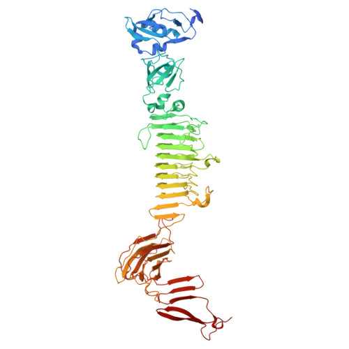
Pantoea stewartii WceF is a glycan biofilm-modifying enzyme with a bacteriophage tailspike-like fold.

Irmscher, T.Roske, Y.Gayk, I.Dunsing, V.Chiantia, S.Heinemann, U.Barbirz, S.

(2021) J Biol Chem 296: 100286-100286

  • DOI: https://doi.org/10.1016/j.jbc.2021.100286
  • Primary Citation of Related Structures:  
    6TGF

  • PubMed Abstract: 

    Pathogenic microorganisms often reside in glycan-based biofilms. Concentration and chain length distribution of these mostly anionic exopolysaccharides (EPS) determine the overall biophysical properties of a biofilm and result in a highly viscous environment. Bacterial communities regulate this biofilm state via intracellular small-molecule signaling to initiate EPS synthesis. Reorganization or degradation of this glycan matrix, however, requires the action of extracellular glycosidases. So far, these were mainly described for bacteriophages that must degrade biofilms for gaining access to host bacteria. The plant pathogen Pantoea stewartii (P. stewartii) encodes the protein WceF within its EPS synthesis cluster. WceF has homologs in various biofilm forming plant pathogens of the Erwinia family. In this work, we show that WceF is a glycosidase active on stewartan, the main P. stewartii EPS biofilm component. WceF has remarkable structural similarity with bacteriophage tailspike proteins (TSPs). Crystal structure analysis showed a native trimer of right-handed parallel β-helices. Despite its similar fold, WceF lacks the high stability found in bacteriophage TSPs. WceF is a stewartan hydrolase and produces oligosaccharides, corresponding to single stewartan repeat units. However, compared with a stewartan-specific glycan hydrolase of bacteriophage origin, WceF showed lectin-like autoagglutination with stewartan, resulting in notably slower EPS cleavage velocities. This emphasizes that the bacterial enzyme WceF has a role in P. stewartii biofilm glycan matrix reorganization clearly different from that of a bacteriophage exopolysaccharide depolymerase.


  • Organizational Affiliation

    Physikalische Biochemie, Universität Potsdam, Potsdam, Germany; Department Theory and Bio-Systems, Max Planck Institute of Colloids and Interfaces, Potsdam, Germany.


Macromolecules
Find similar proteins by:  (by identity cutoff)  |  3D Structure
Entity ID: 1
MoleculeChains Sequence LengthOrganismDetailsImage
Exopolysaccharide biosynthesis proteinA [auth F],
B [auth D],
C [auth E]
736Pantoea stewartii subsp. stewartii DC283Mutation(s): 0 
Gene Names: wceFCKS_2251
UniProt
Find proteins for H3REJ8 (Pantoea stewartii subsp. stewartii DC283)
Explore H3REJ8 
Go to UniProtKB:  H3REJ8
Entity Groups  
Sequence Clusters30% Identity50% Identity70% Identity90% Identity95% Identity100% Identity
UniProt GroupH3REJ8
Sequence Annotations
Expand
  • Reference Sequence
Small Molecules
Ligands 2 Unique
IDChains Name / Formula / InChI Key2D Diagram3D Interactions
PG4
Query on PG4

Download Ideal Coordinates CCD File 
D [auth F],
W [auth D]
TETRAETHYLENE GLYCOL
C8 H18 O5
UWHCKJMYHZGTIT-UHFFFAOYSA-N
EDO
Query on EDO

Download Ideal Coordinates CCD File 
AA [auth D]
BA [auth D]
CA [auth D]
DA [auth D]
E [auth F]
AA [auth D],
BA [auth D],
CA [auth D],
DA [auth D],
E [auth F],
EA [auth D],
F,
FA [auth D],
G [auth F],
GA [auth D],
H [auth F],
HA [auth D],
I [auth F],
IA [auth D],
J [auth F],
JA [auth D],
K [auth F],
KA [auth D],
L [auth F],
LA [auth D],
M [auth F],
MA [auth D],
N [auth F],
NA [auth E],
O [auth F],
OA [auth E],
P [auth F],
PA [auth E],
Q [auth F],
QA [auth E],
R [auth F],
RA [auth E],
S [auth F],
SA [auth E],
T [auth F],
TA [auth E],
U [auth F],
UA [auth E],
V [auth F],
X [auth D],
Y [auth D],
Z [auth D]
1,2-ETHANEDIOL
C2 H6 O2
LYCAIKOWRPUZTN-UHFFFAOYSA-N
Experimental Data & Validation

Experimental Data

  • Method: X-RAY DIFFRACTION
  • Resolution: 2.55 Å
  • R-Value Free: 0.273 
  • R-Value Work: 0.227 
  • R-Value Observed: 0.228 
  • Space Group: P 1 21 1
Unit Cell:
Length ( Å )Angle ( ˚ )
a = 100.179α = 90
b = 97.196β = 106.4
c = 128.949γ = 90
Software Package:
Software NamePurpose
REFMACrefinement
PDB_EXTRACTdata extraction
XDSdata reduction
XSCALEdata scaling
PHASERphasing

Structure Validation

View Full Validation Report


Entry History & Funding Information

Deposition Data


Funding OrganizationLocationGrant Number
German Research Foundation (DFG)GermanyBA 4046/1-2

Revision History  (Full details and data files)

  • Version 1.0: 2020-11-25
    Type: Initial release
  • Version 1.1: 2021-01-27
    Changes: Database references
  • Version 1.2: 2021-07-21
    Changes: Database references
  • Version 1.3: 2024-05-15
    Changes: Data collection, Database references