6TFU

Crystal Structure of EGFR T790M/V948R in Complex with Covalent Pyrrolopyrimidine 14d


Experimental Data Snapshot

  • Method: X-RAY DIFFRACTION
  • Resolution: 2.00 Å
  • R-Value Free: 0.233 
  • R-Value Work: 0.202 
  • R-Value Observed: 0.203 

wwPDB Validation   3D Report Full Report


Ligand Structure Quality Assessment 


This is version 1.2 of the entry. See complete history


Literature

Targeting Her2-insYVMA with Covalent Inhibitors-A Focused Compound Screening and Structure-Based Design Approach.

Lategahn, J.Hardick, J.Grabe, T.Niggenaber, J.Jeyakumar, K.Keul, M.Tumbrink, H.L.Becker, C.Hodson, L.Kirschner, T.Klovekorn, P.Ketzer, J.Baumann, M.Terheyden, S.Unger, A.Weisner, J.Muller, M.P.van Otterlo, W.A.L.Bauer, S.Rauh, D.

(2020) J Med Chem 63: 11725-11755

  • DOI: https://doi.org/10.1021/acs.jmedchem.0c00870
  • Primary Citation of Related Structures:  
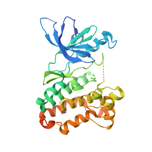
    6TFU, 6TFV, 6TFW, 6TFY, 6TFZ, 6TG0, 6TG1

  • PubMed Abstract: 

    Mutated or amplified Her2 serves as a driver of non-small cell lung cancer or mediates resistance toward the inhibition of its family member epidermal growth factor receptor with small-molecule inhibitors. To date, small-molecule inhibitors targeting Her2 which can be used in clinical routine are lacking, and therefore, the development of novel inhibitors was undertaken. In this study, the well-established pyrrolopyrimidine scaffold was modified with structural motifs identified from a screening campaign with more than 1600 compounds, which were applied against wild-type Her2 and its mutant variant Her2-A775_G776insYVMA. The resulting inhibitors were designed to covalently target a reactive cysteine in the binding site of Her2 and were further optimized by means of structure-based drug design utilizing a set of obtained complex crystal structures. In addition, the analysis of binding kinetics and absorption, distribution, metabolism, and excretion parameters as well as mass spectrometry experiments and western blot analysis substantiated our approach.


  • Organizational Affiliation

    Faculty of Chemistry and Chemical Biology, TU Dortmund University, Otto-Hahn-Strasse 4a, 44227 Dortmund, Germany.


Macromolecules
Find similar proteins by:  (by identity cutoff)  |  3D Structure
Entity ID: 1
MoleculeChains Sequence LengthOrganismDetailsImage
Epidermal growth factor receptor
A, B
333Homo sapiensMutation(s): 2 
Gene Names: EGFRERBBERBB1HER1
EC: 2.7.10.1
UniProt & NIH Common Fund Data Resources
Find proteins for P00533 (Homo sapiens)
Explore P00533 
Go to UniProtKB:  P00533
PHAROS:  P00533
GTEx:  ENSG00000146648 
Entity Groups  
Sequence Clusters30% Identity50% Identity70% Identity90% Identity95% Identity100% Identity
UniProt GroupP00533
Sequence Annotations
Expand
  • Reference Sequence
Experimental Data & Validation

Experimental Data

  • Method: X-RAY DIFFRACTION
  • Resolution: 2.00 Å
  • R-Value Free: 0.233 
  • R-Value Work: 0.202 
  • R-Value Observed: 0.203 
  • Space Group: P 21 21 21
Unit Cell:
Length ( Å )Angle ( ˚ )
a = 76.43α = 90
b = 81.22β = 90
c = 89.92γ = 90
Software Package:
Software NamePurpose
PHENIXrefinement
PDB_EXTRACTdata extraction
XDSdata reduction
XSCALEdata scaling
PHASERphasing

Structure Validation

View Full Validation Report


Ligand Structure Quality Assessment 


Entry History & Funding Information

Deposition Data


Funding OrganizationLocationGrant Number
German Federal Ministry for Education and ResearchGermanyBMBF 01GS08104
German Federal Ministry for Education and ResearchGermanyBMBF 01ZX1303C
European Regional Development FundGermanyEFRE-800400

Revision History  (Full details and data files)

  • Version 1.0: 2020-09-30
    Type: Initial release
  • Version 1.1: 2020-11-04
    Changes: Database references
  • Version 1.2: 2024-01-24
    Changes: Data collection, Database references, Refinement description