Lysozyme crystals dyed with bromophenol blue: where has the dye gone?
Plaza-Garrido, M., Salinas-Garcia, M.C., Alba-Elena, D., Martinez, J.C., Camara-Artigas, A.(2020) Acta Crystallogr D Struct Biol 76: 845-856
- PubMed: 32876060 Search on PubMed
- DOI: https://doi.org/10.1107/S2059798320008803
- Primary Citation Related Structures:
6SYC, 6SYD, 6SYE - PubMed Abstract:
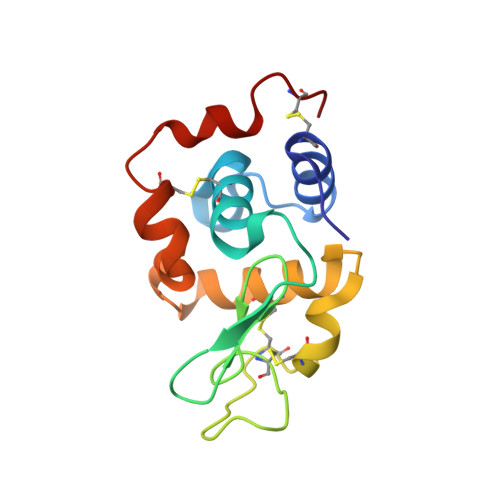
Protein crystals can easily be coloured by adding dyes to their mother liquor, but most structures of these protein-dye complexes remain unsolved. Here, structures of lysozyme in complex with bromophenol blue obtained by soaking orthorhombic and tetragonal crystals in a saturated solution of the dye at different pH values from 5.0 to 7.5 are reported. Two different binding sites can be found in the lysozyme-bromophenol blue crystals: binding site I is located near the amino- and carboxyl-termini, while binding site II is located adjacent to helices α1 (residues 4-15) and α3 (residues 88-100). In the orthorhombic crystals soaked at pH 7.0, binding of the dye takes place in both sites without significant changes in the unit cell. However, soaking tetragonal crystals with bromophenol blue results in two different complexes. Crystals soaked at pH 5.5 (HEWL-T1) show a single dye molecule bound to site II, and the crystals belong to space group P4 3 2 1 2 without significant changes in the unit cell (a = b = 78.50, c = 37.34 Å). On the other hand, crystals soaked at pH 6.5 in the presence of imidazole (HEWL-T2) show up to eight molecules of the dye bound to site II, and display changes in space group (P2 1 2 1 2 1 ) and unit cell (a = 38.00, b = 76.65, c = 84.86 Å). In all of the structures, the dye molecules are placed at the surface of the protein near to positively charged residues accessible through the main solvent channels of the crystal. Differences in the arrangement of the dye molecules at the surface of the protein suggest that the binding is not specific and is mainly driven by electrostatic interactions.
- Department of Chemistry and Physics, Agrifood Campus of International Excellence (ceiA3) and CIAMBITAL, University of Almería, Carretera de Sacramento s/n, 04120 Almería, Spain.
Organizational Affiliation: