6SUR

The Rab33B-Atg16L1 crystal structure


Experimental Data Snapshot

  • Method: X-RAY DIFFRACTION
  • Resolution: 3.47 Å
  • R-Value Free: 0.239 
  • R-Value Work: 0.206 
  • R-Value Observed: 0.207 

wwPDB Validation   3D Report Full Report


Ligand Structure Quality Assessment 


This is version 1.2 of the entry. See complete history


Literature

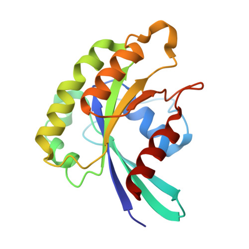
Crystal structure of the Rab33B/Atg16L1 effector complex.

Metje-Sprink, J.Groffmann, J.Neumann, P.Barg-Kues, B.Ficner, R.Kuhnel, K.Schalk, A.M.Binotti, B.

(2020) Sci Rep 10: 12956-12956

  • DOI: https://doi.org/10.1038/s41598-020-69637-0
  • Primary Citation of Related Structures:  
    6SUR

  • PubMed Abstract: 

    The Atg12-Atg5/Atg16L1 complex is recruited by WIPI2b to the site of autophagosome formation. Atg16L1 is an effector of the Golgi resident GTPase Rab33B. Here we identified a minimal stable complex of murine Rab33B(30-202) Q92L and Atg16L1(153-210). Atg16L1(153-210) comprises the C-terminal part of the Atg16L1 coiled-coil domain. We have determined the crystal structure of the Rab33B Q92L/Atg16L1(153-210) effector complex at 3.47 Å resolution. This structure reveals that two Rab33B molecules bind to the diverging α-helices of the dimeric Atg16L1 coiled-coil domain. We mutated Atg16L1 and Rab33B interface residues and found that they disrupt complex formation in pull-down assays and cellular co-localization studies. The Rab33B binding site of Atg16L1 comprises 20 residues and immediately precedes the WIPI2b binding site. Rab33B mutations that abolish Atg16L binding also abrogate Rab33B association with the Golgi stacks. Atg16L1 mutants that are defective in Rab33B binding still co-localize with WIPI2b in vivo. The close proximity of the Rab33B and WIPI2b binding sites might facilitate the recruitment of Rab33B containing vesicles to provide a source of lipids during autophagosome biogenesis.


  • Organizational Affiliation

    Department of Neurobiology, Max-Planck-Institute for Biophysical Chemistry, 37077, Göttingen, Germany. janina.metje@julius-kuehn.de.


Macromolecules
Find similar proteins by:  (by identity cutoff)  |  3D Structure
Entity ID: 1
MoleculeChains Sequence LengthOrganismDetailsImage
Ras-related protein Rab-33B
A, B, C, D, E
A, B, C, D, E, F
173Mus musculusMutation(s): 1 
Gene Names: Rab33b
UniProt & NIH Common Fund Data Resources
Find proteins for O35963 (Mus musculus)
Explore O35963 
Go to UniProtKB:  O35963
IMPC:  MGI:1330805
Entity Groups  
Sequence Clusters30% Identity50% Identity70% Identity90% Identity95% Identity100% Identity
UniProt GroupO35963
Sequence Annotations
Expand
  • Reference Sequence
Find similar proteins by:  (by identity cutoff)  |  3D Structure
Entity ID: 2
MoleculeChains Sequence LengthOrganismDetailsImage
Autophagy-related protein 16-157Mus musculusMutation(s): 0 
Gene Names: Atg16l1Apg16l
UniProt & NIH Common Fund Data Resources
Find proteins for Q8C0J2 (Mus musculus)
Explore Q8C0J2 
Go to UniProtKB:  Q8C0J2
IMPC:  MGI:1924290
Entity Groups  
Sequence Clusters30% Identity50% Identity70% Identity90% Identity95% Identity100% Identity
UniProt GroupQ8C0J2
Sequence Annotations
Expand
  • Reference Sequence
Small Molecules
Ligands 2 Unique
IDChains Name / Formula / InChI Key2D Diagram3D Interactions
GTP
Query on GTP

Download Ideal Coordinates CCD File 
N [auth A]
P [auth B]
R [auth C]
T [auth D]
V [auth E]
N [auth A],
P [auth B],
R [auth C],
T [auth D],
V [auth E],
X [auth F]
GUANOSINE-5'-TRIPHOSPHATE
C10 H16 N5 O14 P3
XKMLYUALXHKNFT-UUOKFMHZSA-N
MG
Query on MG

Download Ideal Coordinates CCD File 
M [auth A]
O [auth B]
Q [auth C]
S [auth D]
U [auth E]
M [auth A],
O [auth B],
Q [auth C],
S [auth D],
U [auth E],
W [auth F]
MAGNESIUM ION
Mg
JLVVSXFLKOJNIY-UHFFFAOYSA-N
Experimental Data & Validation

Experimental Data

  • Method: X-RAY DIFFRACTION
  • Resolution: 3.47 Å
  • R-Value Free: 0.239 
  • R-Value Work: 0.206 
  • R-Value Observed: 0.207 
  • Space Group: P 1 21 1
Unit Cell:
Length ( Å )Angle ( ˚ )
a = 48.4α = 90
b = 204.9β = 92.6
c = 107.2γ = 90
Software Package:
Software NamePurpose
PHENIXrefinement
Cootmodel building
PHASERphasing
XSCALEdata scaling
XDSdata reduction

Structure Validation

View Full Validation Report


Ligand Structure Quality Assessment 


Entry History & Funding Information

Deposition Data


Funding OrganizationLocationGrant Number
German Research FoundationGermanySFB860

Revision History  (Full details and data files)

  • Version 1.0: 2020-08-05
    Type: Initial release
  • Version 1.1: 2020-08-12
    Changes: Database references
  • Version 1.2: 2024-01-24
    Changes: Data collection, Database references, Refinement description